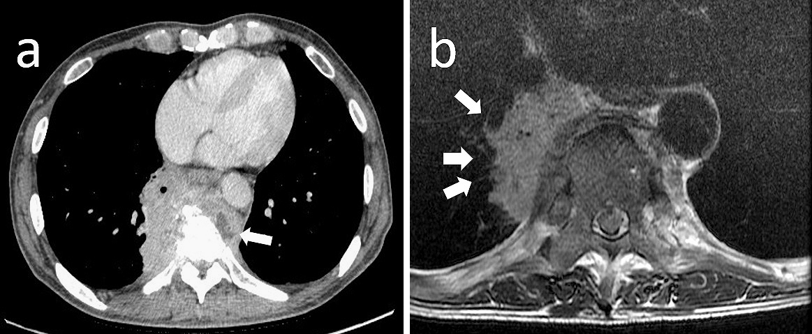
Figure 1. Imaging studies on admission.
(a) Contrast-enhanced computed tomography (CT) on admission demonstrated an invasive mass lesion with non-homogenous enhancement. The mass measured 85 × 67 mm and was located adjacent to S7 of the right lung, with bone infiltration and a compression fracture of the thoracic vertebrae. A low-density area (white arrow) was partially visible on the left side of the mass. (b) A magnetic resonance (MR) T2-weighted image showed that the mass spiraled up the pulmonary vessels (white arrows) and markedly infiltrated the thoracic vertebrae.

From: A Case Report of an Intrathoracic Mass Lesion Caused by Arcanobacterium haemolyticum that Required Exclusion of a Malignant Tumor Diagnosis

Figure 2. Biopsy findings.
A computed tomography-guided biopsy specimen demonstrated inflammatory granulation by neutrophils, histiocytes, plasma cells, and lymphocytes, which was nonspecific inflammation in the subacute and chronic phase. Hematoxylin & eosin stain, magnification × 50.

From: A Case Report of an Intrathoracic Mass Lesion Caused by Arcanobacterium haemolyticum that Required Exclusion of a Malignant Tumor Diagnosis

Figure 3. Results of tissue and blood cultures.
(a) β-hemolysis on sheep blood agar; (b) Gram stain of Arcanobacterium haemolyticum in blood culture.

From: A Case Report of an Intrathoracic Mass Lesion Caused by Arcanobacterium haemolyticum that Required Exclusion of a Malignant Tumor Diagnosis

PAGE TOP