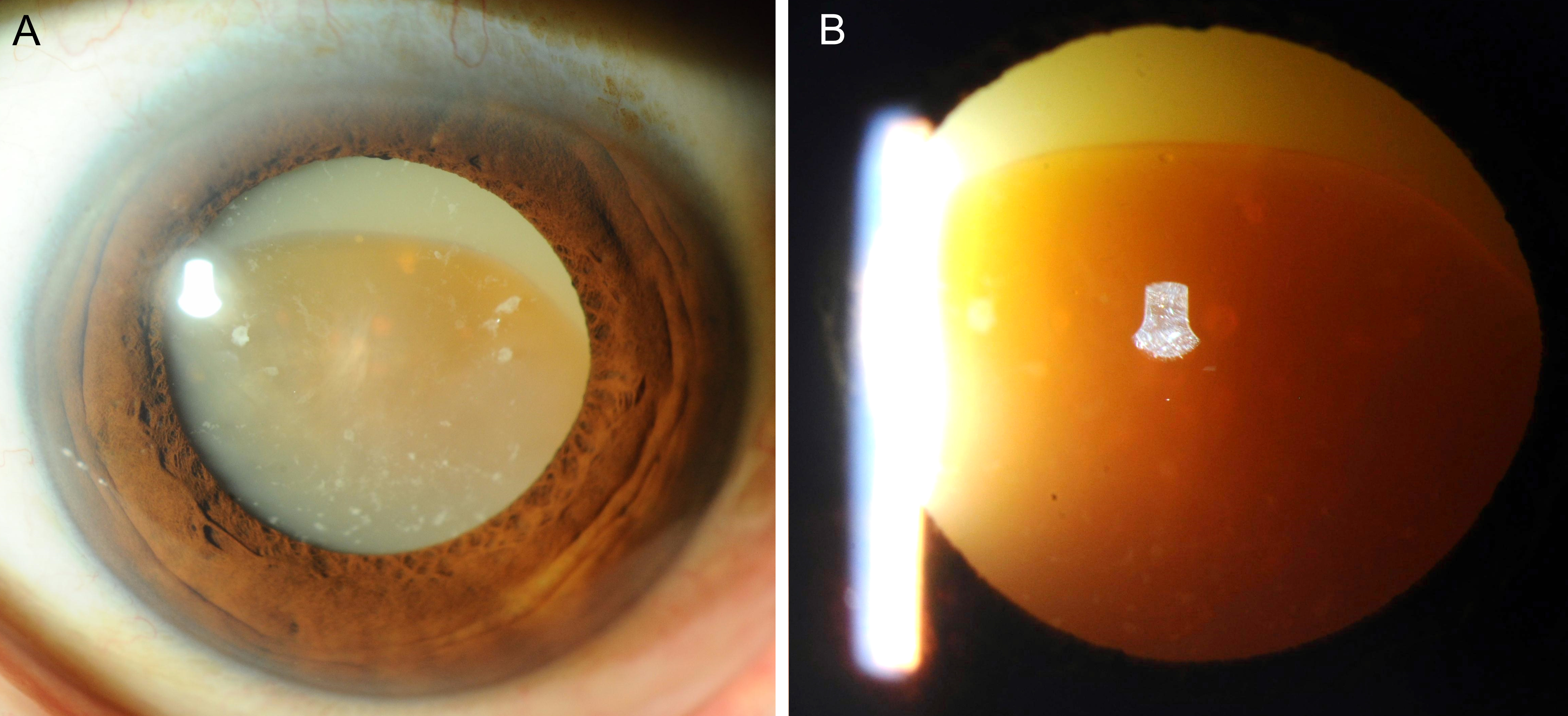
Figure 1. A. Image of the anterior segment obtained using slit-lamp microscopy at initial presentation. Morgagnian cataract is observed. The cortex is emulsified and the brown nucleus is sinking. B. Image of the anterior eye segment with transillumination. The solid nucleus of the lens and total liquefaction of the cortex can be observed.
From: Morgagnian Cataract