Figure 1. Brain computed tomography findings.
A. Axial nonenhanced computed tomography (NECT) scan shows typical findings of pineal germinoma with a well-demarcated slightly hyperdense tumor engulfing the calcified pineal gland.
B. Sagittal NECT scan shows a lobulated hyperdense mass with different locations, suprasellar, infundibular, and tumor spread, in the lateral ventricles and third ventricle with hydrocephalus.
From: Type D Adipsia with Severe Hypernatremia: A Unique Presentation of an Extensive Intracranial Germinoma
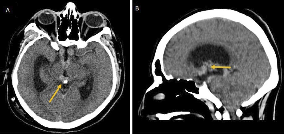
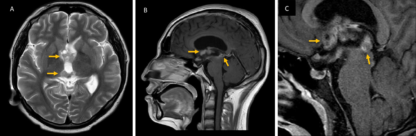
Figure 2. Brain magnetic resonance imaging (MRI) findings.
A. T2WI MRI Axial Lesions common isointense and hyperintense in the third ventricle with multiple cysts in the germinoma. B. Sagittal T1WI C+ MRI shows homogeneous suprasellar enhancement of the hypothalamus and chiasmatic region, with extension to the pineal gland through the floor of the third ventricle. C. Sagital T1WI C MR. Large suprasellar lesion with obstructive hydrocephalus with the appearance of an enlarged lateral ventricle and flattening of the fornix.
From: Type D Adipsia with Severe Hypernatremia: A Unique Presentation of an Extensive Intracranial Germinoma