Corresponding author: Junki Mizumoto, jnk_mizu@yahoo.co.jp
DOI: 10.31662/jmaj.2023-0178
Received: November 8, 2023
Accepted: December 18, 2023
Advance Publication: February 9, 2024
Published: April 15, 2024
Cite this article as:
Otsubo S, Mizumoto J. Idiopathic Iliopsoas Muscle Hematoma. JMA J. 2024;7(2):288-289.
Key words: Emergency Medicine, Hematoma, Iliopsoas muscle
A 77-year-old man complained of severe right lateral back pain. It had developed acutely nine days earlier when the patient was riding bicycle on a steep incline; the pain relieved seven days earlier. After cycling again on the day before presentation, the pain recurred. On the day of the presentation, the pain suddenly worsened.
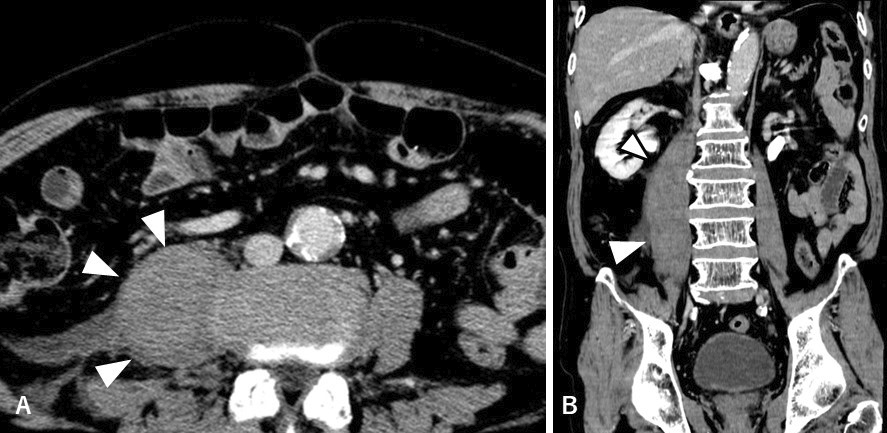
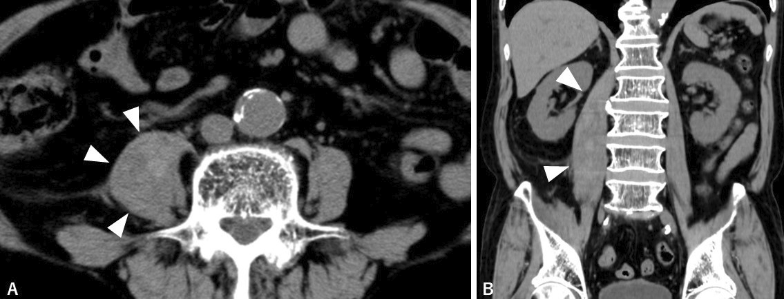
He had to lay down on the bed and could not move his lower limbs; he remained afebrile. Computed tomography revealed distended right iliopsoas muscle (Figure 1). There was no contrast enhancement within the lump or along its periphery, reducing the likelihood of an iliopsoas abscess. Considering the homogeneously contrasted lump and abrupt onset of pain, idiopathic iliopsoas hematoma was ascertained to be the most likely diagnosis. Laboratory tests revealed no coagulopathies. Shrinkage of the iliopsoas lump was confirmed on the fourth day after the admission (Figure 2), concomitant with pain relief, leading to a definitive diagnosis.
This case highlights that iliopsoas hematomas can develop in patients without predisposing factors (1), and even moderate mechanical stress can trigger their development (2). Furthermore, the pain progressing in a stepwise manner suggests a potential link between repetitive strain and hematoma development.
None
Concept: S.O., J.M.; design: S.O., J.M.; data collection or processing: S.O., J.M.; analysis or interpretation: S.O., J.M.; literature review: S.O., J.M.; and writing: S.O., J.M.
Informed consent was obtained from the patient.
Shinya Otsubo: 0009-0007-0689-653X
Junki Mizumoto: 0000-0002-0783-7351
Marquardt G, Barduzal Angles S, Leheta F, et al. Spontaneous haematoma of the iliac psoas muscle: a case report and review of the literature. Arch Orthop Trauma Surg. 2002;122(2):109-11.
Kawarat V, Javid M, Swaminathan SP, et al. A rare case of iliopsoas hematoma in a patient with von Willebrand disease. Int Surg J. 2020;7(3):873-5.
