Corresponding author: Keisuke Watanabe, YCUmedRDCkw@yahoo.co.jp, watanabk@yokohama-cu.ac.jp
DOI: 10.31662/jmaj.2025-0030
Received: January 18, 2025
Accepted: May 22, 2025
Advance Publication: August 1, 2025
Key words: computed tomography, congenital disorders, unilateral absence of the pulmonary artery
A 55-year-old woman was referred to our hospital owing to dyspnea on exertion and abnormalities on chest images. Her vital signs were unremarkable. Physical examination revealed normal vascular sounds, no murmurs, no jugular vein distention, and no edema. Chest radiography showed decreased size of the right hemithorax, elevation of the right diaphragm, and absence of the right pulmonary artery shadow (Figure 1). Chest computed tomography (CT) showed absence of the right pulmonary artery and right lung volume loss (Figure 2). The echocardiogram showed no congenital heart disease and no pulmonary hypertension. The patient was diagnosed with congenital isolated absence of the right pulmonary artery.
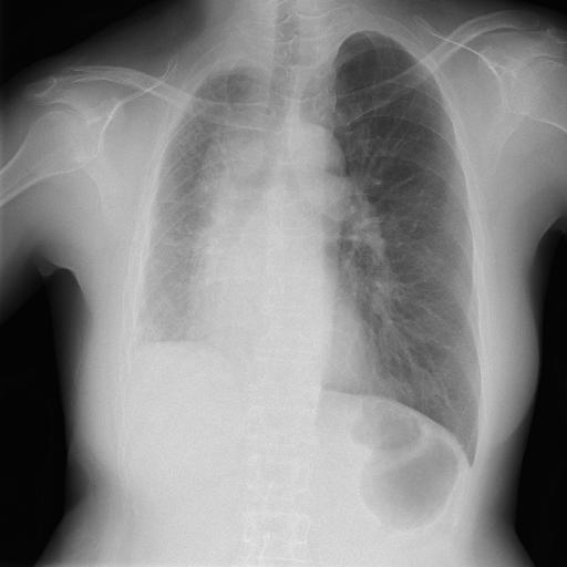
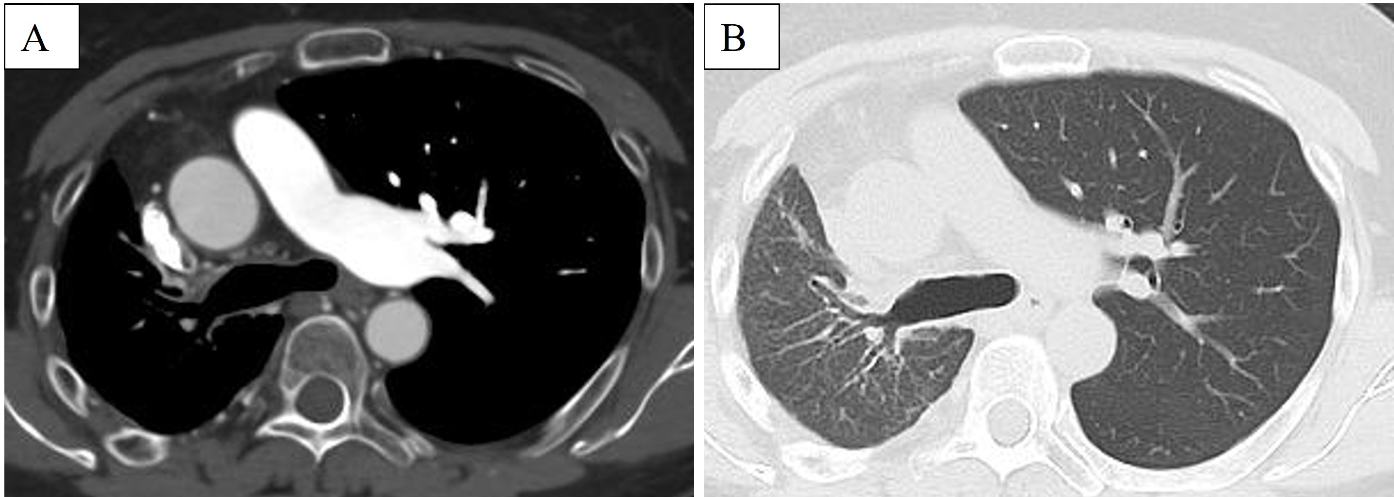
The features of chest radiography in this disease include a decreased size of the affected hemithorax, an absent ipsilateral, and enlarged contralateral pulmonary artery shadow (1), (2). Chest CT shows a unilateral absence of the pulmonary artery, volume loss of the affected lung, and a systemic-to-pulmonary shunt (1), (2).
None
Keisuke Watanabe collected the clinical data and wrote the initial draft of the manuscript. Takeshi Kaneko supervised and edited the manuscript. Both authors read and approved the final manuscript.
This study was approved by the institutional review board of Yokohama City University Hospital (number 2024-041).
Written informed consent was obtained from the patient to publish the case report.
During the preparation of this work, the authors used ChatGPT to improve readability and language. After using this tool/service, the authors reviewed and edited the content as needed and take full responsibility for the content of the publication.
Jariwala P, Maturu VN, Christopher J, et al. Congenital isolated unilateral agenesis of pulmonary arteries in adults: case series and review. Indian J Thorac Cardiovasc Surg. 2021;37(suppl 1):144-54.
Wang P, Yuan L, Shi J, et al. Isolated unilateral absence of pulmonary artery in adulthood: a clinical analysis of 65 cases from a case series and systematic review. J Thorac Dis. 2017;9(12):4988-96.
