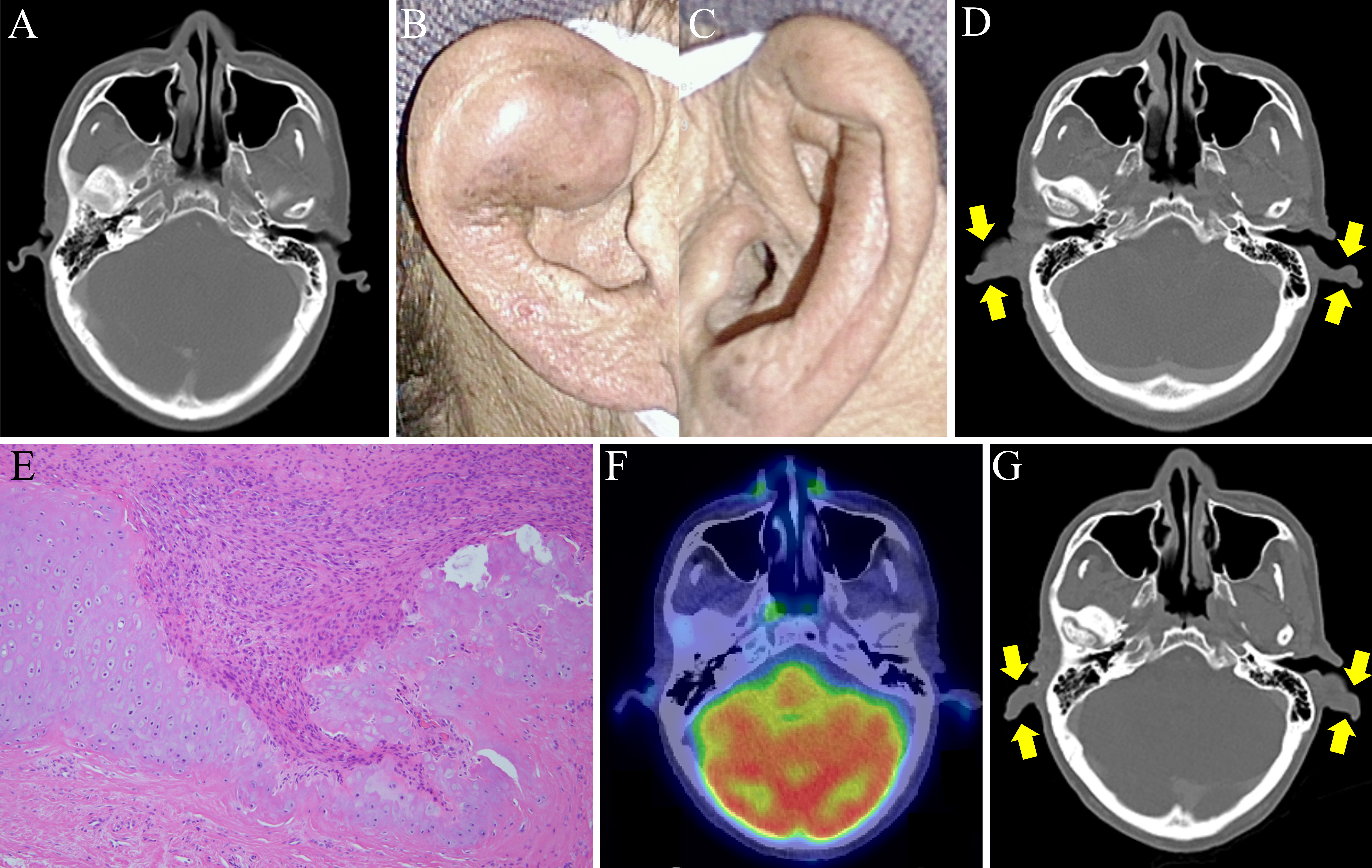
Figure 1. Auricular involvement in a patient with relapsing polychondritis induced by pembrolizumab, a programmed cell death protein-1 inhibitor.
(A) No enlargement in the bilateral auricles was observed in CT before pembrolizumab therapy. (B-D) Swelling of the bilateral auricles developed 4 months after pembrolizumab therapy (B, right ear; C, left ear; D, yellow arrows in CT indicate auricular involvement). (E) Hematoxylin-eosin-stained images of the right auricle reveal cartilage destruction due to infiltration of the surrounding granulation tissue (magnification, 100×). (F) Lack of 18F-FDG uptake in the regressed swelling of bilateral auricles on 18F-FDG-PET/CT 1 month after interruption of pembrolizumab therapy. (G) CT showing relapsing auricular involvement after three cycles of pembrolizumab therapy. Yellow arrows indicate the reswelling of the bilateral auricles. 18F-FDG-PET/CT, 18F-fluorodeoxyglucose-positron emission tomography/computed tomography.
From: Relapsing Polychondritis following PD-1 Blockade by an Immune Checkpoint Inhibitor