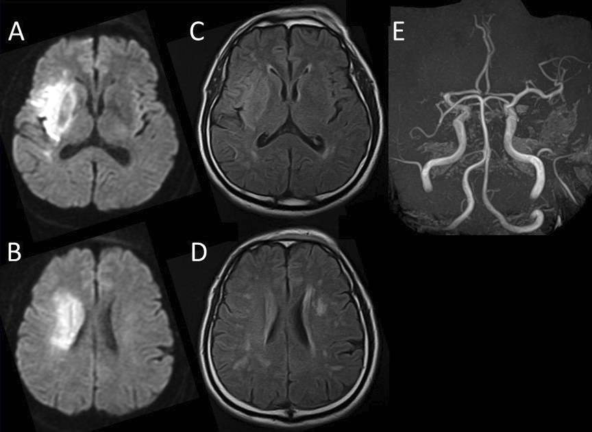
Figure 1. Initial magnetic resonance imaging A, B: diffusion-weighted imaging, C, D: fluid-attenuated inversion recovery and magnetic resonance angiography (E) acute infarction in the right middle cerebral artery (MCA) territory and MCA occlusion.
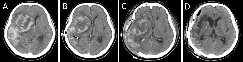
Figure 2. Head computed tomography (CT) indicated hemorrhagic infarction within a known cerebral infarct, midline shift, and cerebral herniation (A). Postoperative head CT indicated a progressively less midline shift after small craniotomy (B), right frontotemporal large decompressive craniectomy (C), and cranioplasty (D).
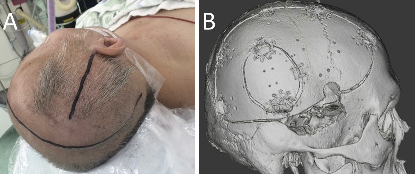
Figure 3. Photograph shows skin incision lines during small craniotomy and large decompressive craniectomy (A). Three-dimensional computed tomography image after cranioplasty (B).