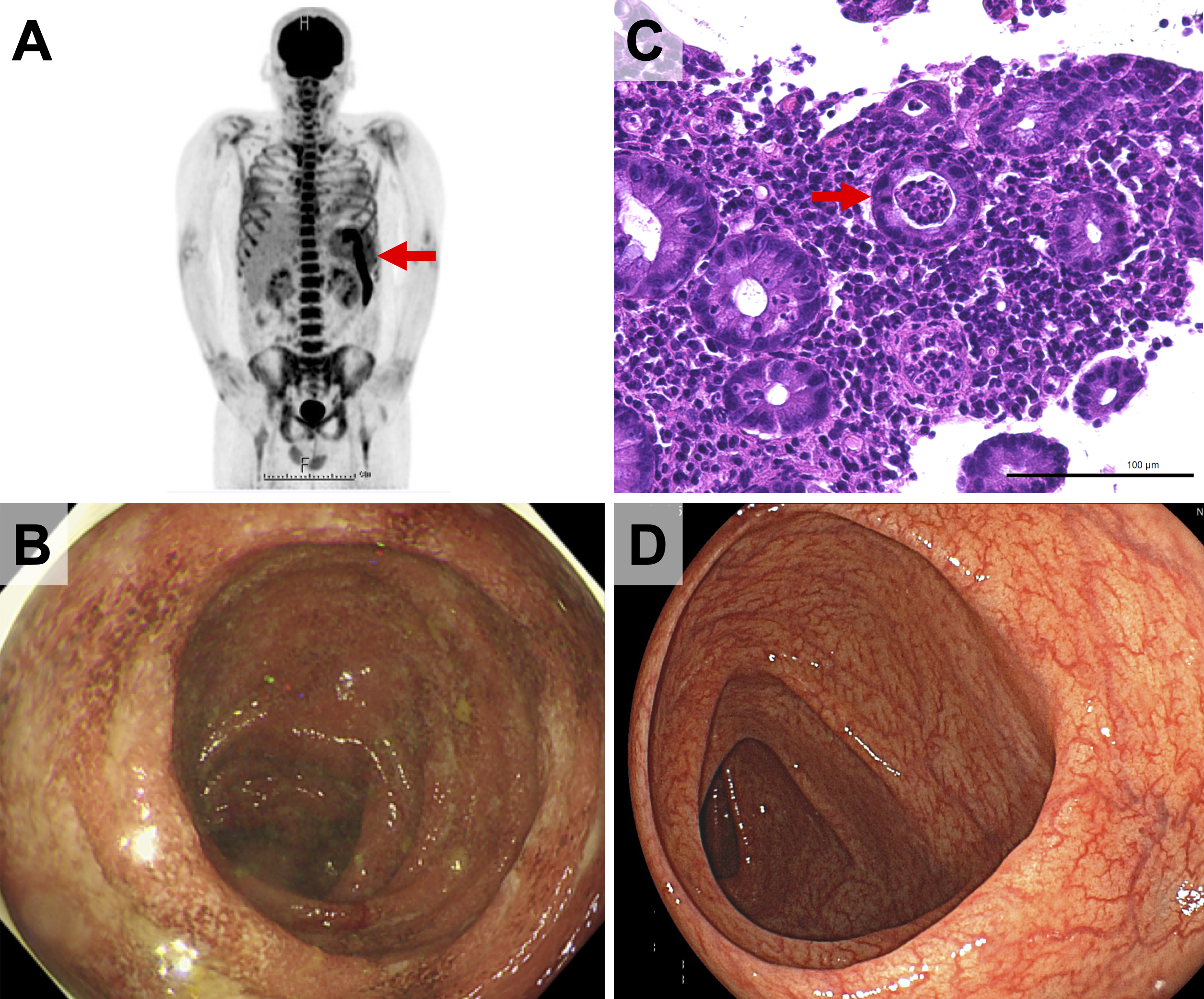
Figure 1. (A) 18F-fluorodeoxyglucose positron emission tomography and computed tomography revealing high uptake in the spleen, bone marrow, systemic lymph nodes, and splenic flexure of the colon (arrow). (B) Colonoscopic images showing mucosal friability, erosions, and multiple shallow ulcers in the area from the transverse colon to the rectum. (C) Histological evaluation revealing dense mucosal inflammatory infiltration and crypt abscesses (arrow). (D) Four-month follow-up colonoscopy showing the improvement of the colitis.
From: Bloody Diarrhea in a 27-year-old Man with Adult-onset Still’s Disease