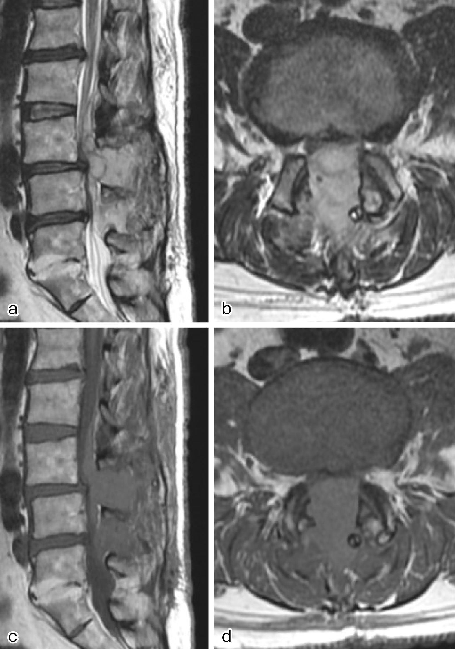
Figure 2. Several hours after the initial surgery, magnetic resonance imaging (MRI) was performed to investigate the cause of bilateral lower limb paralysis and pain. MRI revealed the presence of an epidural hematoma. (a) Sagittal and (b) horizontal sections of T2-weighted images, and (c) sagittal and (d) horizontal sections of T1-weighted images.
From: A Case of Epidural Hematoma after Lumbar Spine Surgery in a Hemophilia B Carrier