Figure 1. Illustration of the AI support in echocardiography workflows
View Classification―AI identifies and labels the different views of the echocardiographic images. View Classification―AI classifies the images into specific types of echocardiographic views, such as apical 4-chamber (4ch). Auto Measurements―AI performs automatic measurements of cardiac structures, such as left ventricular end-diastolic volume (LVEDV). Echo AI Report―AI generates comprehensive echocardiographic reports based on automated measurements and analyses. AI Diagnosis―AI assists in diagnosing cardiac conditions by analyzing the echocardiographic data.

From: AI in Echocardiography: State-of-the-art Automated Measurement Techniques and Clinical Applications

Figure 2. Comparison between conventional and automated strain measurements in echocardiography
Conventional measurement involves manual steps like chamber selection and ROI modification (left). Automated measurement (right) requires no manual intervention and provides instant results. ROI: region of interest.

From: AI in Echocardiography: State-of-the-art Automated Measurement Techniques and Clinical Applications

Figure 3. Impact of the image quality on the measurement time in echocardiography
The left side shows examples of good-quality images compared to fair- or poor-quality images. The right-side graph demonstrates that fair- or poor-quality images require significantly more time for measurements (p < 0.01) compared to good-quality images, while reporting time remains consistent (ns).

From: AI in Echocardiography: State-of-the-art Automated Measurement Techniques and Clinical Applications

Figure 4. Erroneous automated measurement case of the posterior wall using AI
Inclusion of chordae tendineae in the measurement results in a wall thickness overestimation (16.4 mm), potentially leading to a misdiagnosis of concentric hypertrophy. This case highlights the necessity for an accurate anatomical identification in AI-driven echocardiography.

From: AI in Echocardiography: State-of-the-art Automated Measurement Techniques and Clinical Applications

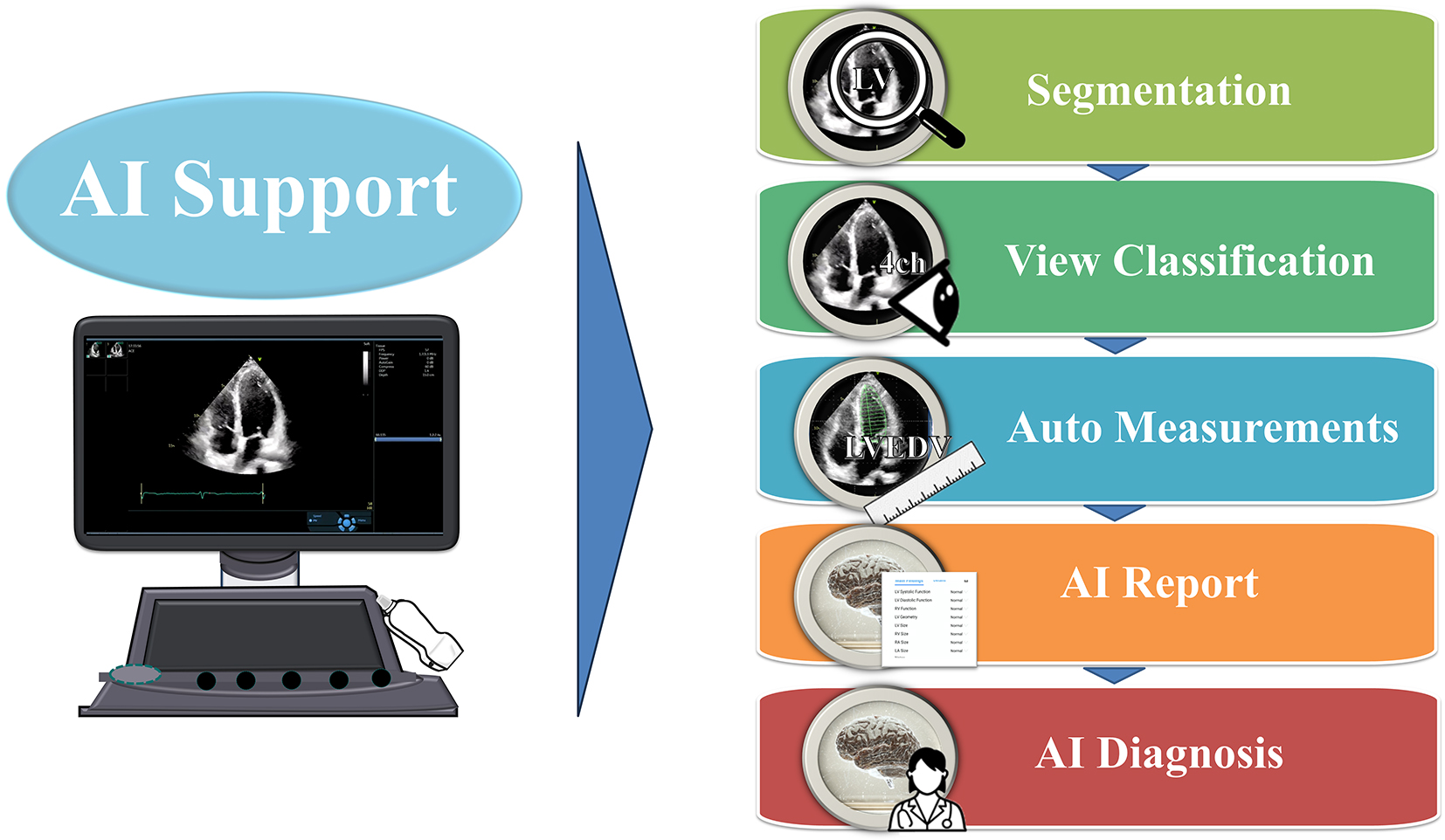
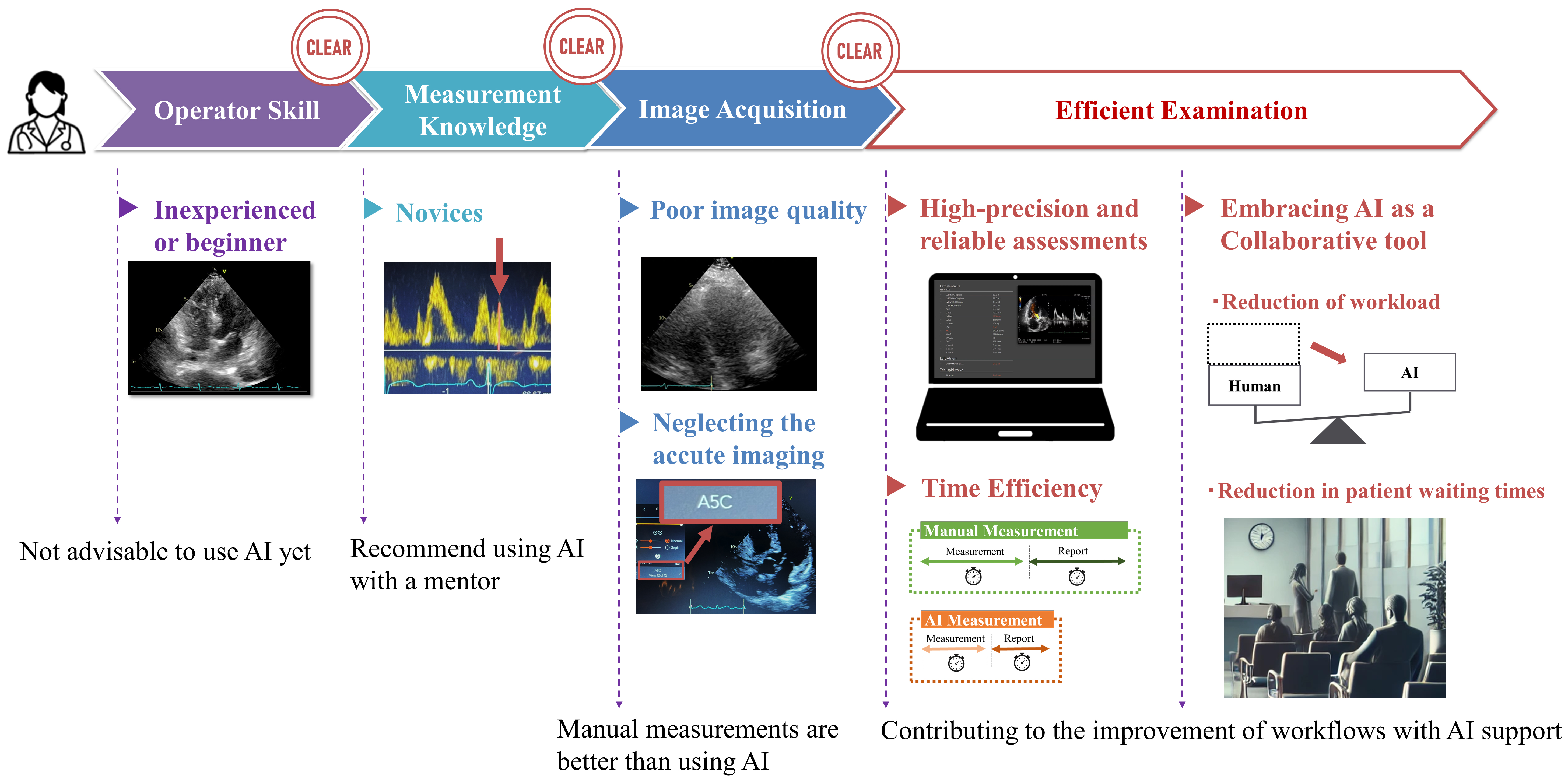
Figure 5. Strategies for the effective use of AI in echocardiography
Inexperienced operators should not use AI alone, while novices are advised to use AI with a mentor. Manual measurements are better with a poor image quality or a neglected imaging. Once these challenges are addressed, high-precision and reliable assessments, improved time efficiency, and reduced workload and patient waiting times are achieved by integrating AI as a collaborative tool.

From: AI in Echocardiography: State-of-the-art Automated Measurement Techniques and Clinical Applications

PAGE TOP