Figure 1. Picture of the patient’s face showing bilateral periorbital purpura.

From: Periorbital Purpura as a Key Diagnostic Clue in Immunoglobulin Light Chain Amyloidosis

Figure 2. Biopsy specimen of rectal mucosa showing amorphous extracellular material with positive Congo red staining.

From: Periorbital Purpura as a Key Diagnostic Clue in Immunoglobulin Light Chain Amyloidosis
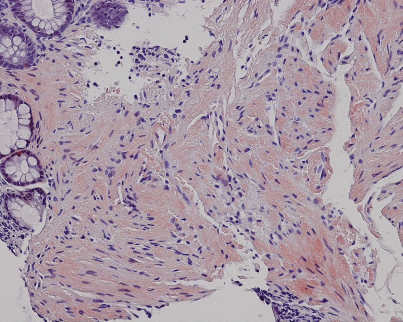
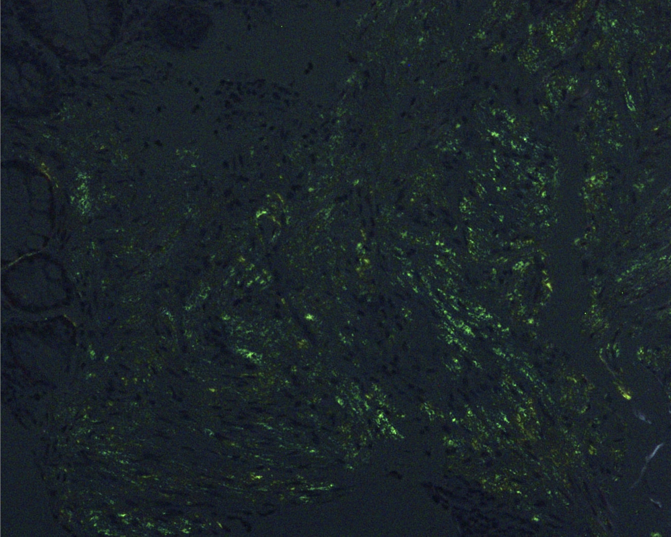
Figure 3. Biopsy specimen of rectal mucosa showing apple-green birefringence under polarized light microscopy.

From: Periorbital Purpura as a Key Diagnostic Clue in Immunoglobulin Light Chain Amyloidosis
PAGE TOP