Home
Advance Publications
Issues
Instructions for Authors
Guidelines
Submission
Editors & Publishers
Acknowledgement
JMA Journal
Home
Advance Publications
Issues
Instructions for Authors
Guidelines
Submission
Editors & Publishers
Acknowledgement
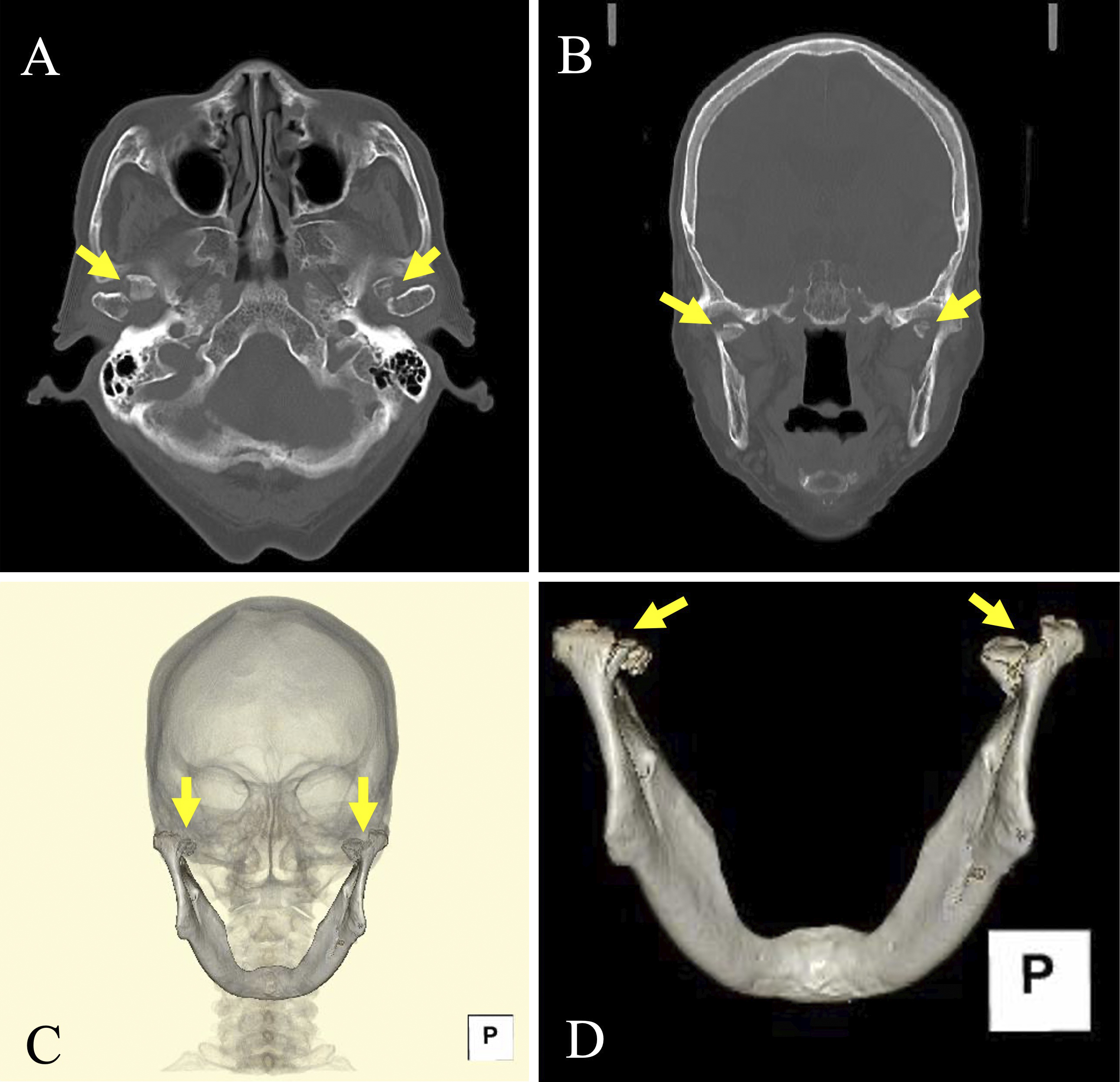
Figure 1.
Computed tomography images. (A) Axial image. (B) Coronal image. (C) Three-dimensional (3D)-reconstructed image. (D) Mandibular 3D-reconstructed image. Arrows indicate fracture lines.
From:
Bilateral Mandibular Condylar Fractures
» Back to article page
©
Japan Medical Association
. All rights reserved.