Figure 1. (A) Clinical photograph of Case 1. A black nodule on the left chest, with surrounding pigmented patches. (B) Pathological findings of Case 1. A dense proliferation of atypical melanocytes was seen in the lesion (Hematoxylin-eosin staining, original magnification, ×200). (C) Magnetic resonance imaging before the treatment with pembrolizumab and stereotactic radiotherapy in Case 1. Multiple brain metastases were seen (white arrows). (D) Magnetic resonance imaging 5 years after the treatment with pembrolizumab and stereotactic radiotherapy in Case 1.
From: Two Cases of Malignant Melanoma with Long-term Survival after the Appearance of Brain Metastases
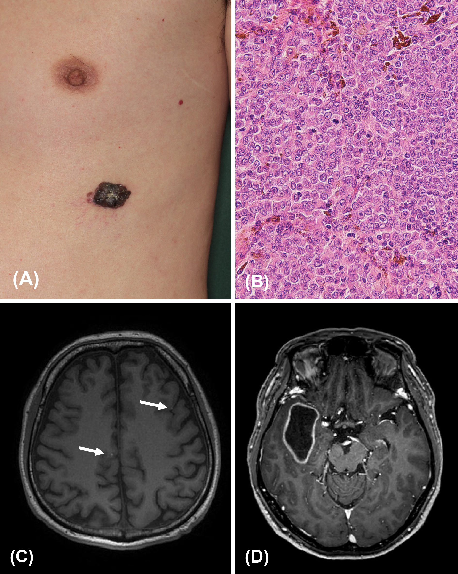
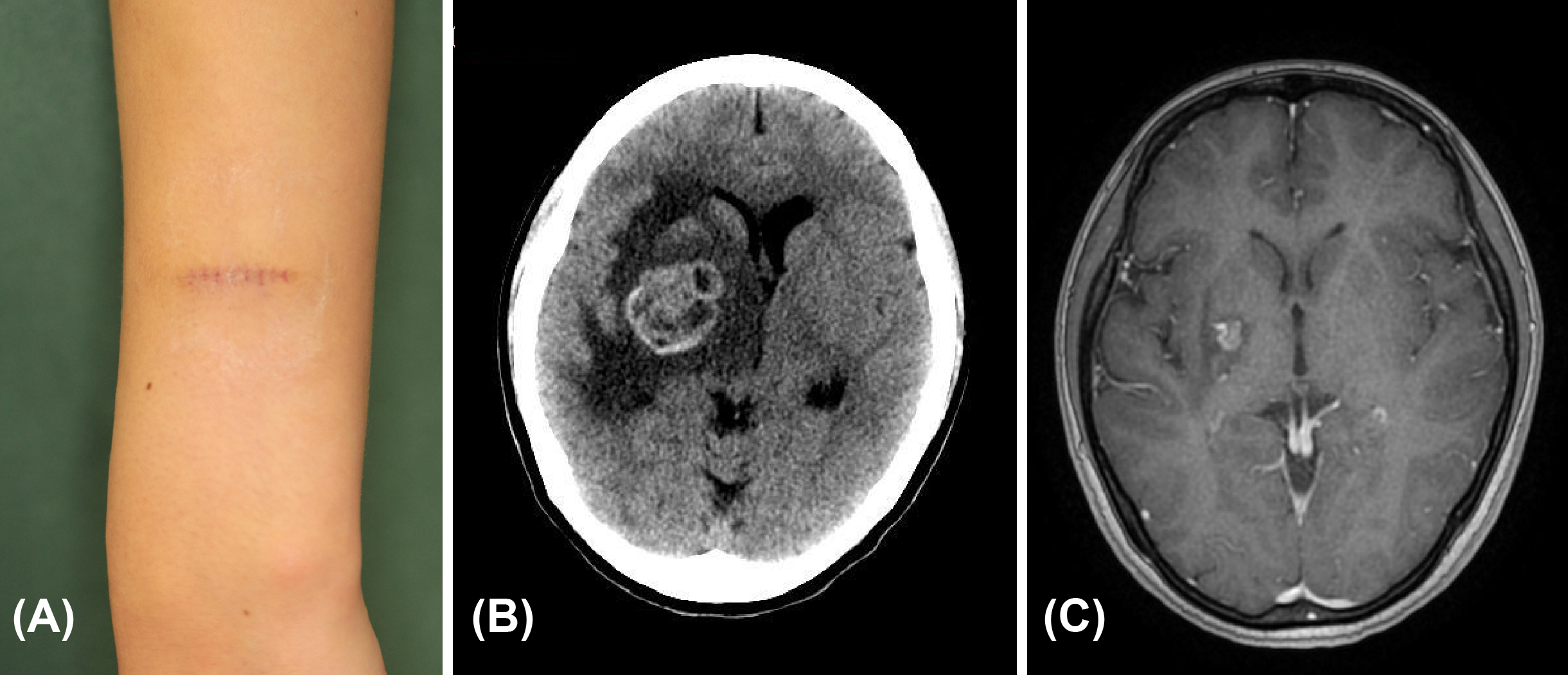
Figure 2. (A) Clinical photograph of Case 2. There were no residual lesions around the scar. (B) Computerized tomography before the treatment with chemotherapy and stereotactic radiotherapy in Case 2. Metastasis measuring 35 mm in diameter is seen in the right basal ganglia. (C) Magnetic resonance imaging 4 years after the treatment with pembrolizumab and stereotactic radiotherapy in Case 2. The size of metastasis has been significantly reduced.
From: Two Cases of Malignant Melanoma with Long-term Survival after the Appearance of Brain Metastases