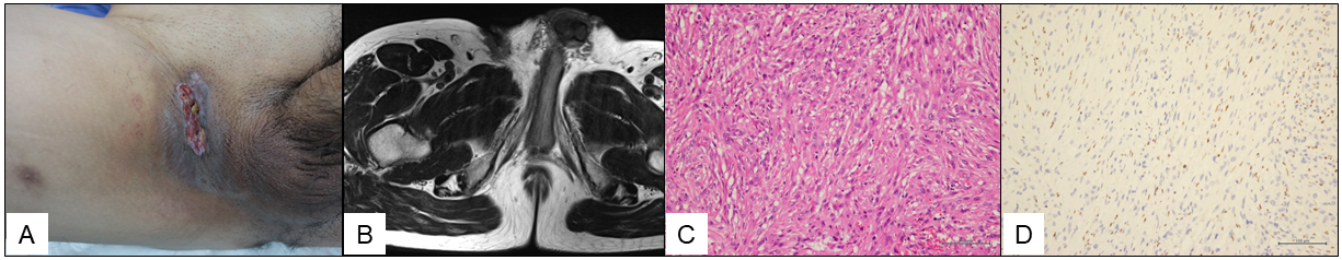
Figure 1. (A) A painless ulcer with firm edges on the right groin, measuring 6 cm × 2 cm. (B) Axial T1-weighted image showed a homogeneous superficial soft-tissue mass infiltrating into the right groin. (C) Large, ovoid epithelioid cells with rich eosinophilic cytoplasm (HE staining). (D) Complete loss of integrase interactor 1 protein expression in the tumor cells.
HE: hematoxylin and eosin; T1: type 1.
From: Epithelioid Sarcoma Mimicking Mycotic Granulomatosis