Figure 2. Histology of the newborn cochlea of the common marmoset. The newborn cochlea of the common marmoset has 2 and a half turns, the same as the adult cochlear (A). Immunohistochemical analysis can be applied to this primate cochlea. One row of inner hair cells and 3 rows of outer hair cells can be clearly observed (B and C). IHC: inner hair cells, MYO7A: hair cell markers; OHC: outer hair cells; SOX2: supporting cell markers.
From: The Common Marmoset as a Novel Non-human Primate Model for Inner Ear Research
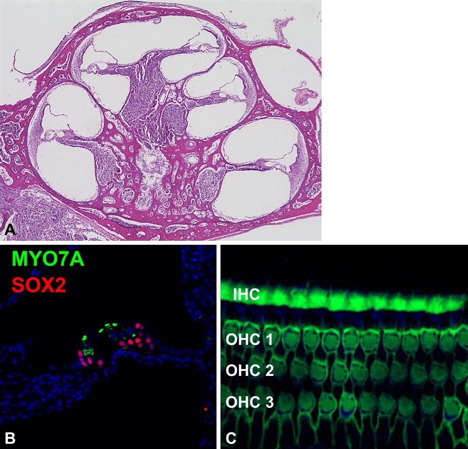
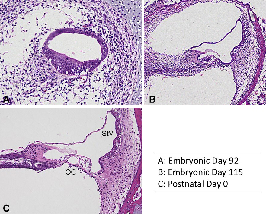
Figure 3. Developmental, morphological changes of the cochlea of the common marmoset. During the cochlear development, the cochlear duct changes its morphology. Previous studies on developmental investigation in primate cochlea have described their general time course. The common marmoset has become a novel developmental study model animal in this field. OC: organ of Corti, StV: stria vascularis.
From: The Common Marmoset as a Novel Non-human Primate Model for Inner Ear Research