Figure 1. (A) Anteroposterior three-dimensional computed tomography angiography (3D-CTA) showed the internal carotid artery (ICA) medially to the external carotid artery (ECA). (B) CTA showed 74% ICA stenosis according to North American Symptomatic Carotid Endarterectomy Trial criteria. (C) The angle of twisting was measured in the axial view on CTA by computing the degree of medial deviation from the line connecting the centers of the ICA and ECA to the line parallel to the sagittal plane at the center of the ECA. The preoperative twist angle was 102°. (D) Fluid-attenuated inversion recovery imaging showed a chronic cerebral infarction in the right parietal lobe. (E) Black-blood T1-weighted image showed a high-intensity signal, indicating an unstable vulnerable plaque. (F) Postoperative anteroposterior 3D-CTA showed improvement of the ICA stenosis without repositioning.
From: Carotid Endarterectomy for a Case with an Extremely Twisted Internal Carotid Artery
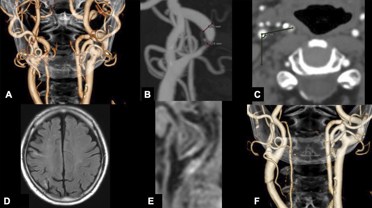
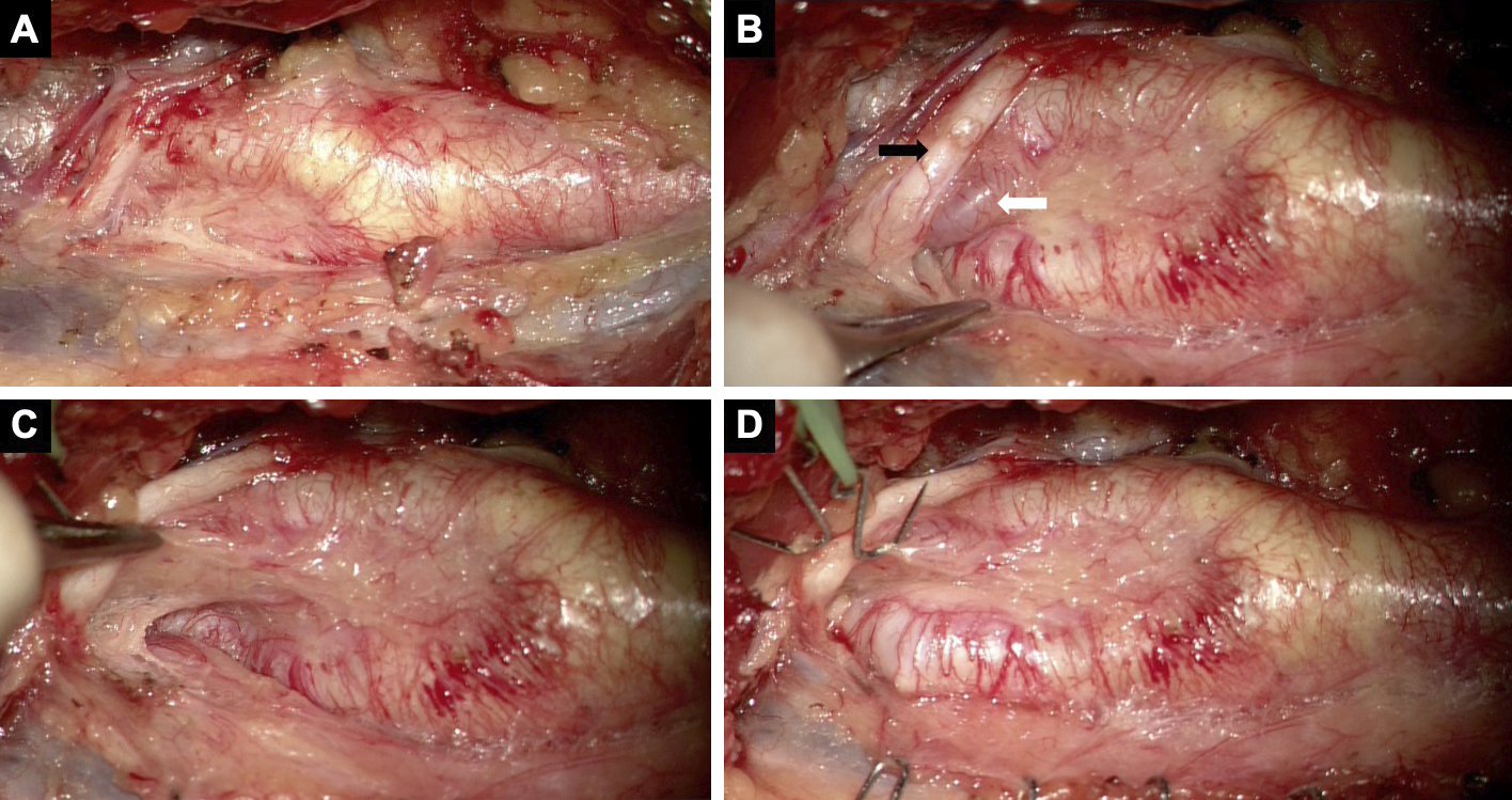
Figure 2. Operative view of the case. (A) The intraoperative photograph at the level of the carotid bifurcation showed the concealed ICA. (B) The ICA was located under the hypoglossal nerve (black arrow) and occipital artery (white arrow). (C) After mobilizing the hypoglossal nerve, the distal ICA ran deeply. (D) After transposing the ICA to its normal location, this surgical field resembled a conventional CEA.
From: Carotid Endarterectomy for a Case with an Extremely Twisted Internal Carotid Artery