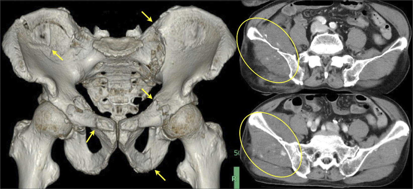
Figure 1. Computed tomography (CT) of the pelvis revealed a pelvic fracture classified as LC-III according to the Young-Burgess classification (arrow). Contrast-enhanced CT also revealed contrast medium extravasation in the pelvic muscles (circle).
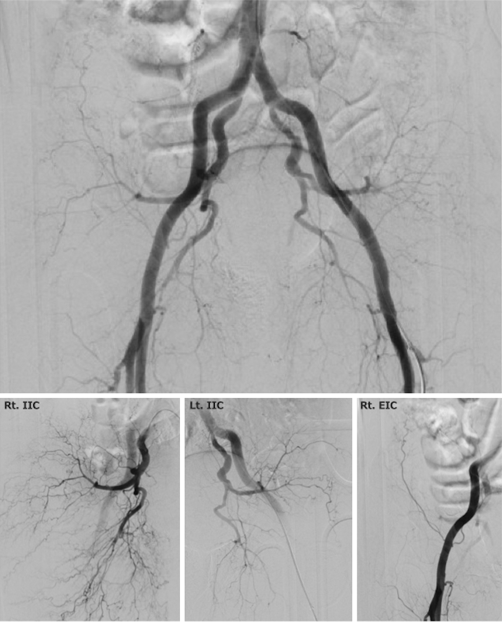
Figure 2. Emergency angiography of the pelvis revealed the disappearance of contrast medium extravasation. Selective angiography revealed that the bleeding was halted. Rt. IIC: right internal iliac artery, Lt. IIC: left internal iliac artery, Rt. EIC: right external iliac artery.