Figure 1. Magnetic resonance angiography of the intracranial arteries at the time of diagnosis.

From: Severe Chronic Mesenteric Ischemia in a Patient with Moyamoya Disease
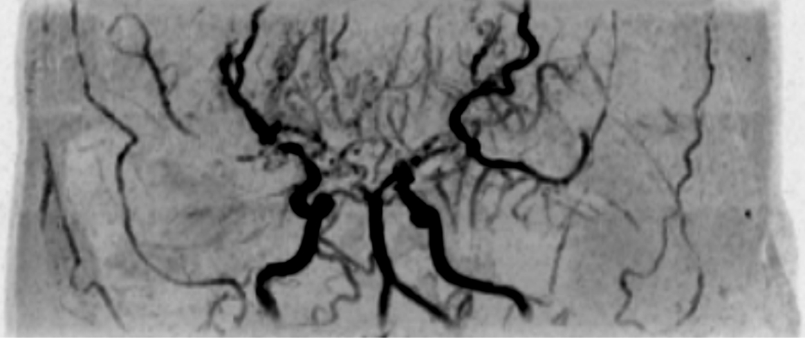
Figure 2. Computed tomography images showing occlusion of the common trunk of the celiac and superior mesenteric arteries (arrow) and the development of prominent collateral arteries via the inferior mesenteric artery. (a) Maximum intensity projection image. (b) Three-dimensional reconstruction image.

From: Severe Chronic Mesenteric Ischemia in a Patient with Moyamoya Disease
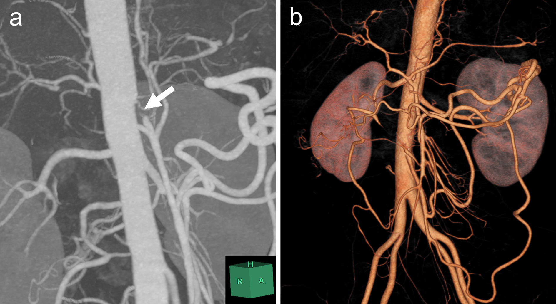
Figure 3. Intraoperative image showing the aorta (arrow) to superior mesenteric artery (arrowhead) bypass using a great saphenous vein graft.

From: Severe Chronic Mesenteric Ischemia in a Patient with Moyamoya Disease
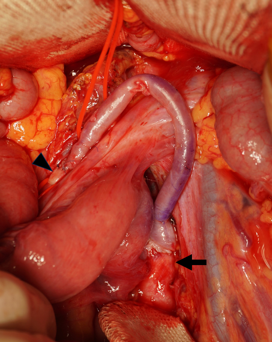
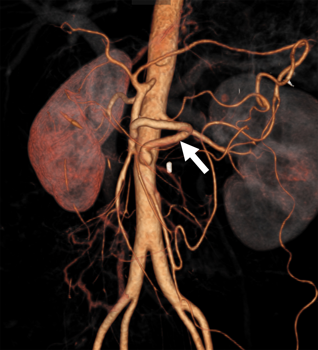
Figure 4. Postoperative computed tomography showing an aorta to superior mesenteric artery bypass (arrow).

From: Severe Chronic Mesenteric Ischemia in a Patient with Moyamoya Disease
PAGE TOP