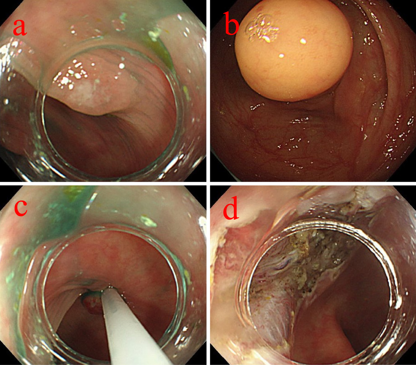
Figure 1. a. The 5 mm Is polyp was found in the transverse colon. b. the 30 mm submucosal tumor was found in the descending colon. c. the 5 mm Is polyp in the transverse colon was resected by CSP. d. ESD was then performed on the 30 mm submucosal tumor in the descending colon.

From: Delayed Perforation after Colorectal Cold Snare Polypectomy with Simultaneously Performed Endoscopic Submucosal Dissection: A Case Report and Literature Review

Figure 2. a. Computed tomography showed free air on the liver surface. b. We identified a 5 × 7 mm perforation in the transverse colon.

From: Delayed Perforation after Colorectal Cold Snare Polypectomy with Simultaneously Performed Endoscopic Submucosal Dissection: A Case Report and Literature Review

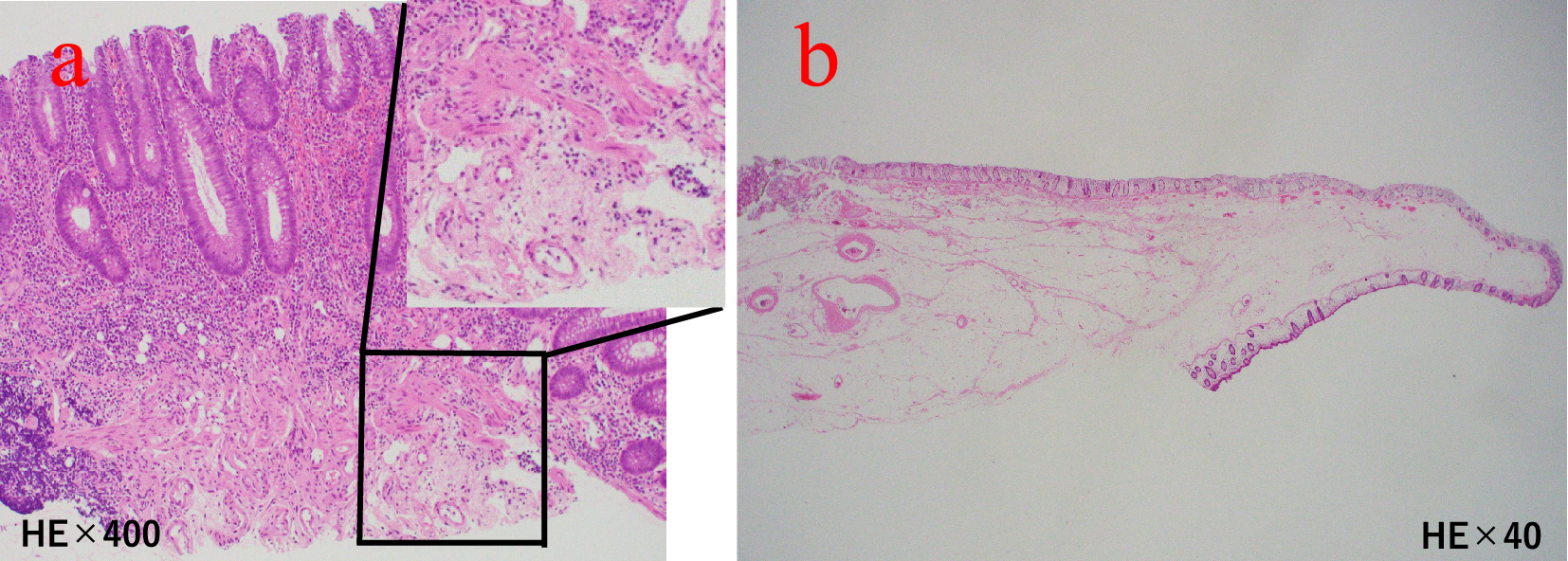
Figure 3. a. Pathological image of a transverse colon polyp is shown. The muscularis propria was not included (hematoxylin and eosin [H&E] stain, ×40 and ×400). b. Pathological image of a descending colon in the submucosal tumor is shown. The muscularis propria was not included in either case (H&E stain, ×40).

From: Delayed Perforation after Colorectal Cold Snare Polypectomy with Simultaneously Performed Endoscopic Submucosal Dissection: A Case Report and Literature Review

PAGE TOP