Home
Advance Publications
Issues
Instructions for Authors
Guidelines
Submission
Editors & Publishers
Acknowledgement
JMA Journal
Home
Advance Publications
Issues
Instructions for Authors
Guidelines
Submission
Editors & Publishers
Acknowledgement
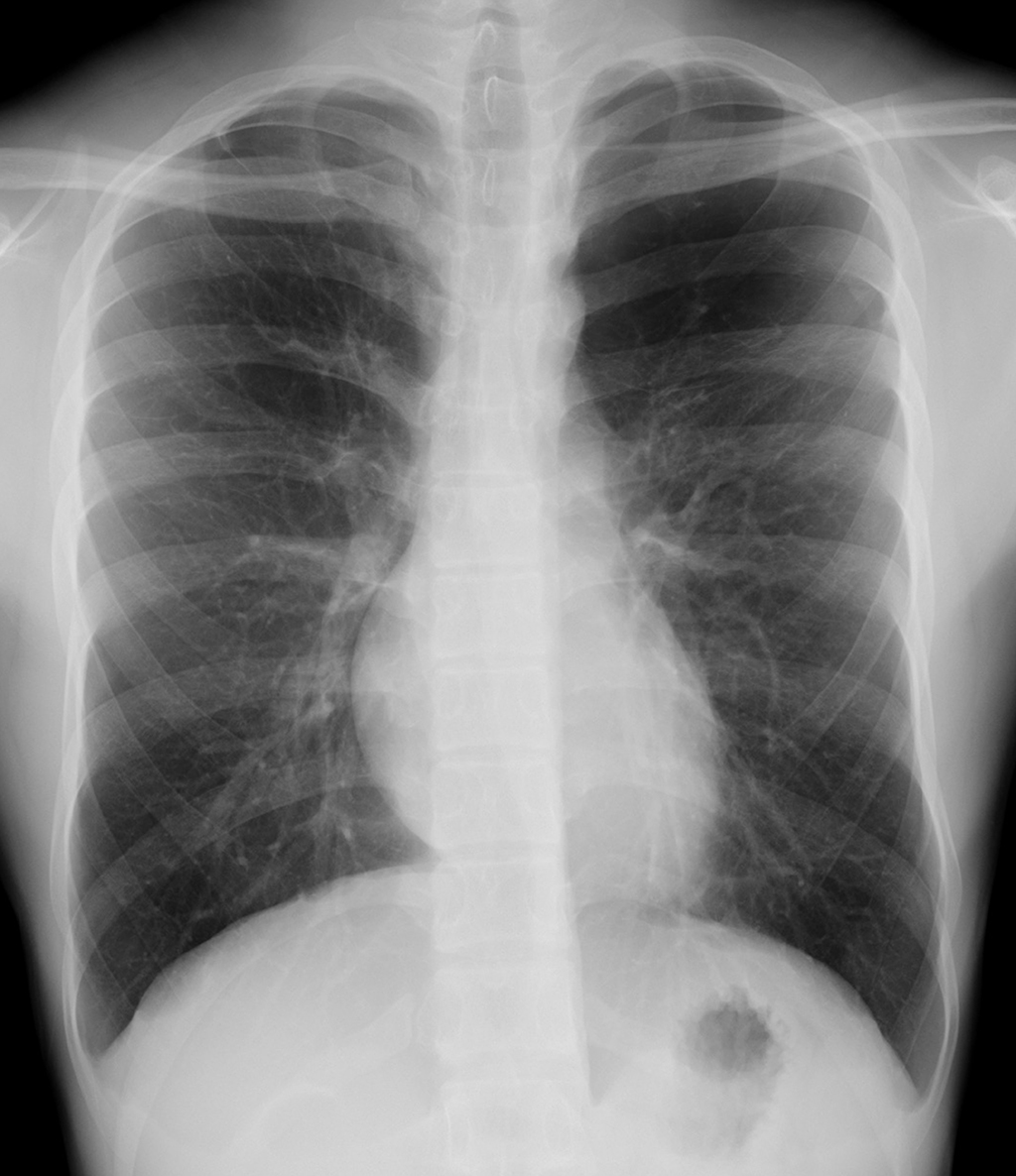
Figure 1.
Chest radiograph showing increased radiolucency in the left upper lung field.
From:
A Case of Asymptomatic Juvenile Emphysema
» Back to article page
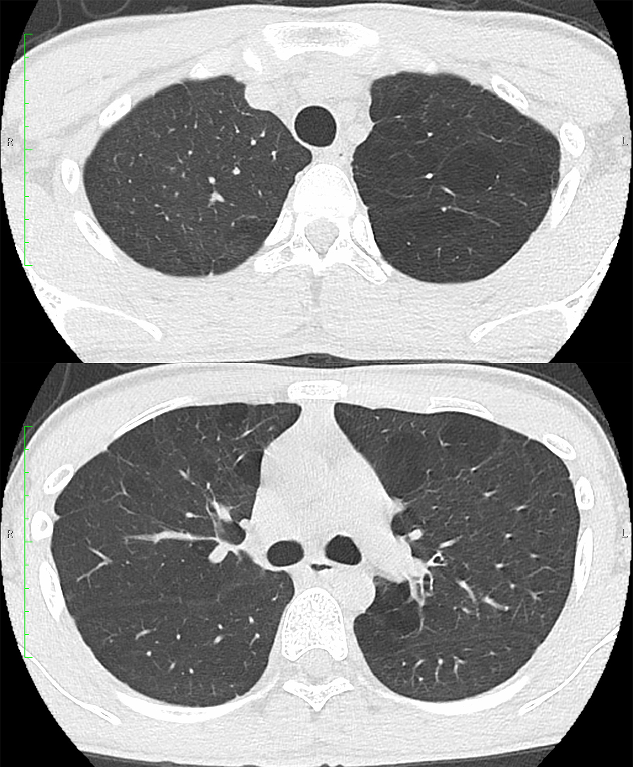
Figure 2.
Chest computed tomography scan showing scattered panlobular emphysema predominantly in the left upper lobe.
From:
A Case of Asymptomatic Juvenile Emphysema
» Back to article page
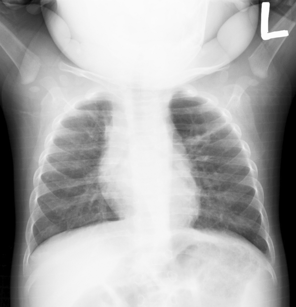
Figure 3.
Chest radiograph at the age of 1 year showing similar increased radiolucency in the left upper lung field.
From:
A Case of Asymptomatic Juvenile Emphysema
» Back to article page
©
Japan Medical Association
. All rights reserved.