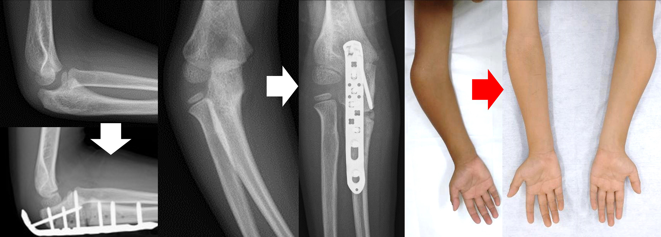
Figure 2. Preoperative planning and intraoperative macrophotography
Osteotomy was performed at the center of rotation of angulation (CORA), slightly distal to the coronoid process of the ulna. An open wedge osteotomy was performed to correct the deformity considering the balance with the radial length. Plate fixation with artificial bone was planned. A corrective osteotomy of 30° was performed as planned while using external fixation.
From: A Pediatric Case of Cubitus Varus Deformity due to Olecranon Fracture
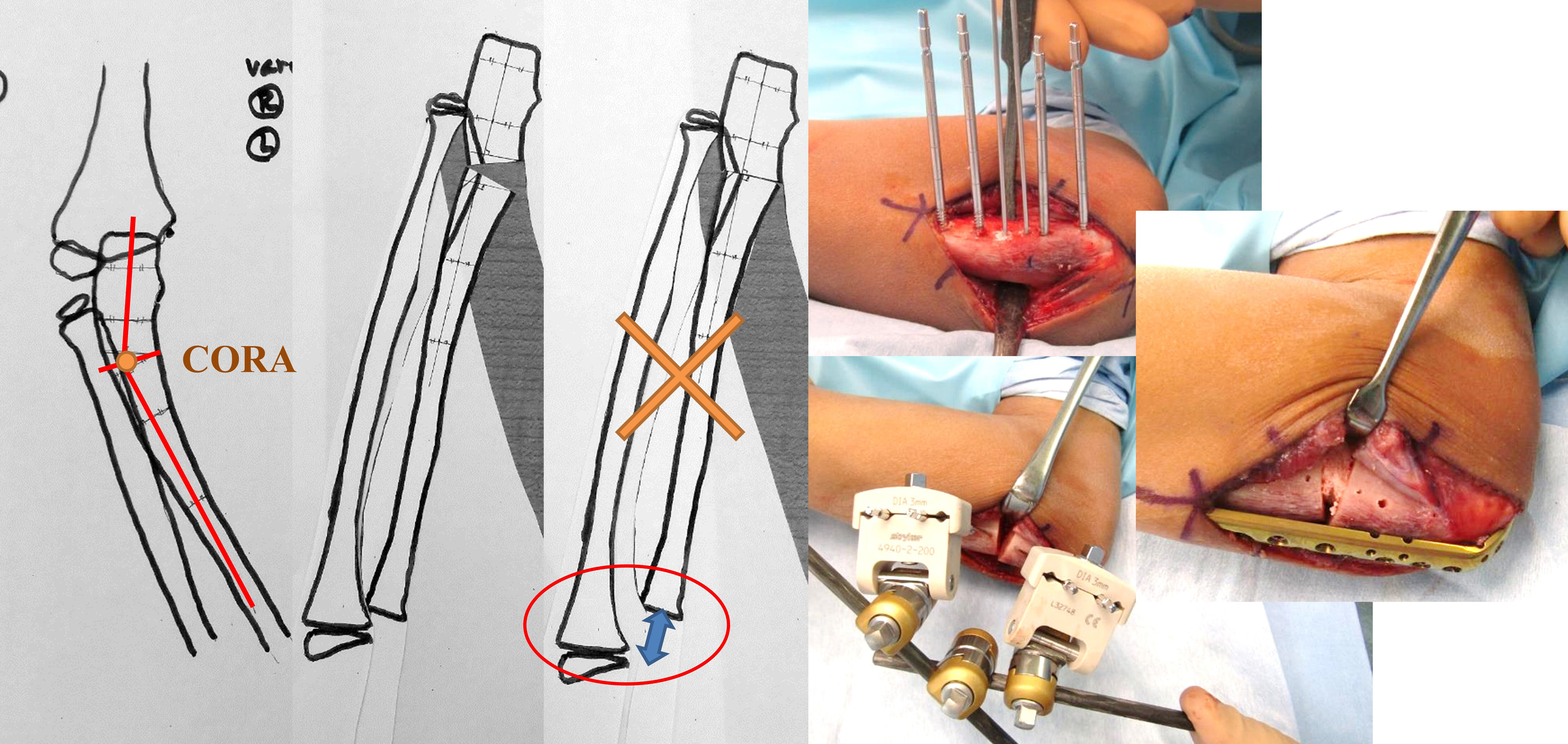
Figure 4. X-ray image at last follow-up
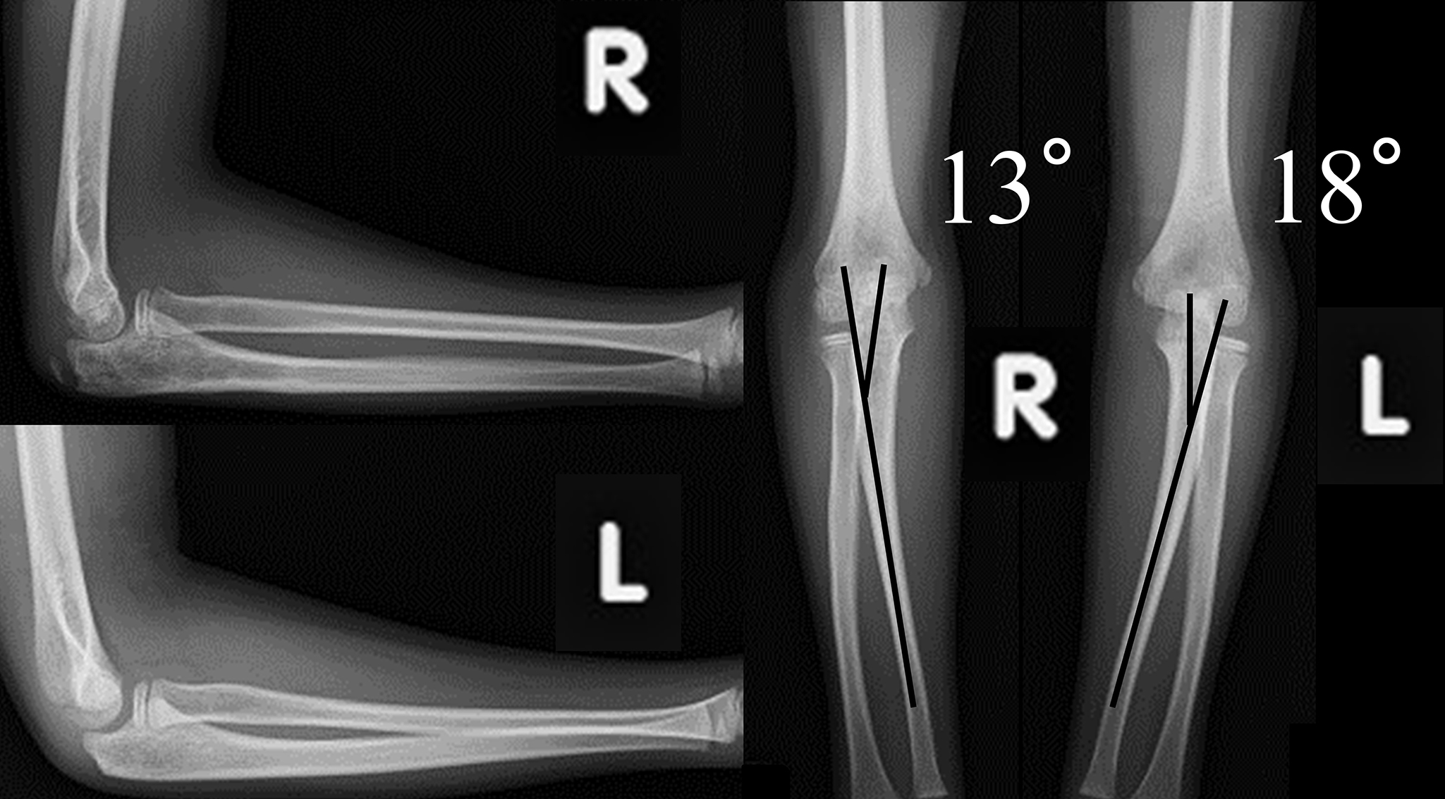
The plate was extracted one year after the corrective osteotomy; the final follow-up was 3.5 years after the injury and 2.5 years after the corrective osteotomy. VA 13° (contralateral side 18°) and CA 10° varus (contralateral side 12° rotation) with little difference between right and left and no limitation of range of motion or pain. VA, varus angulation; CA, carrying angle.
From: A Pediatric Case of Cubitus Varus Deformity due to Olecranon Fracture