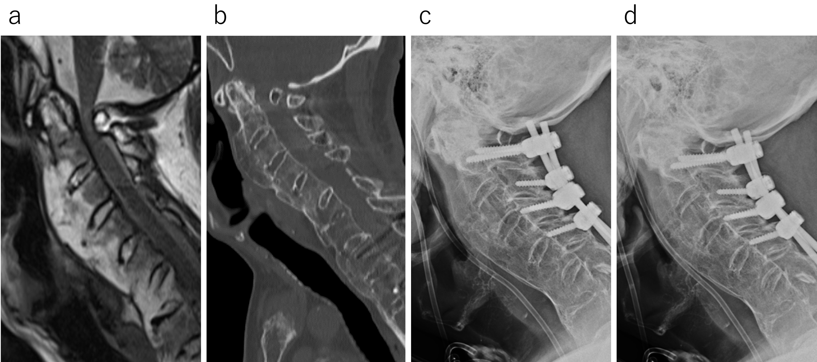
Figure 1. Imaging tests are recommended for the initial consultation. Sagittal T2-weighted MRI demonstrates substantial spinal cord compression at C1/2 (a). The atlantodental interval measures approximately 1 mm on the sagittal CT scan (b). X-ray images of anteflexion (c) and retroflexion (d) similarly reveal minimal instability at C1/2.
Figure 2. Approximately 1 month postsurgery, subcutaneous swelling was noted, prompting an MRI evaluation. The imaging revealed a pseudomeningocele measuring 14 cm in length on the sagittal plane (a) and 4 cm in width on the horizontal plane (b).
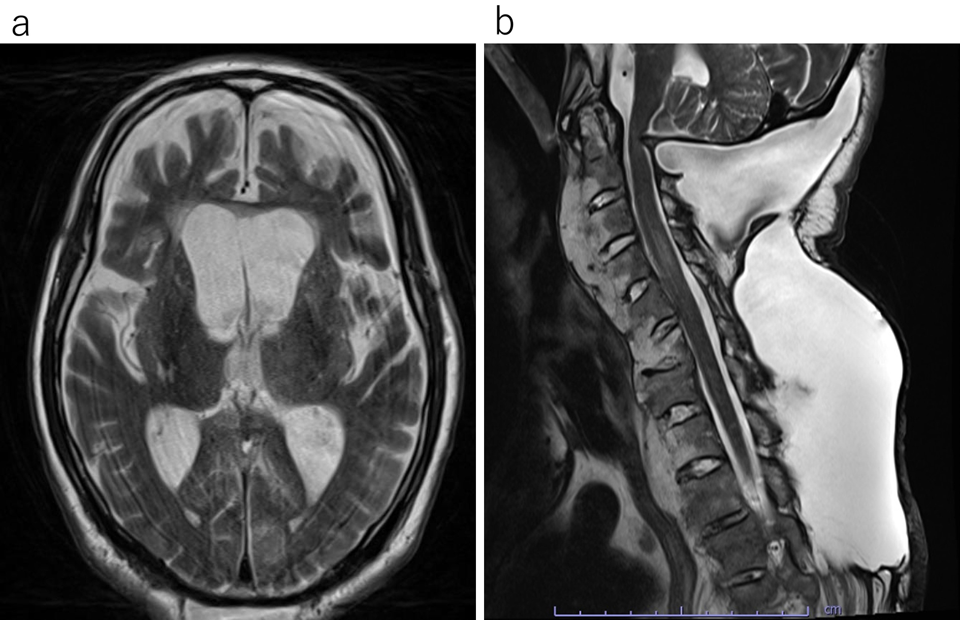
Figure 3. Four months postsurgery, the patient exhibited drowsiness and vomiting. Head and cervical MRIs were performed in response to these symptoms. The head MRI revealed considerable ventricular enlargement (a), whereas the cervical MRI indicated an increase in the pseudomeningocele (b).
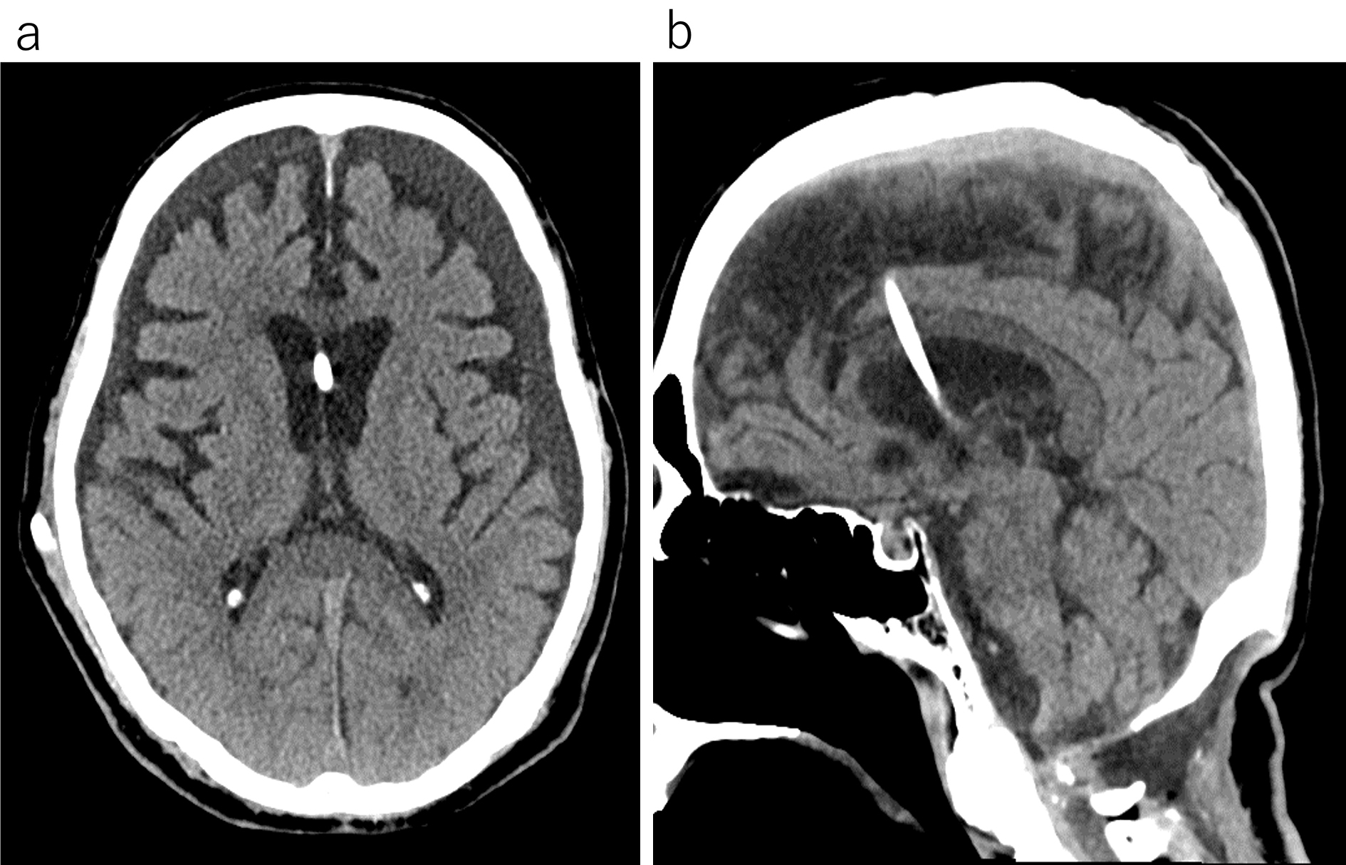
Figure 4. This computed tomography scan of the head, performed after ventriculoperitoneal shunt surgery, shows that the ventricles have returned to normal size (a) and the pseudomeningocele has significantly decreased (b).