Figure 2. High-resolution computed tomography findings. (A) mild peripheral reticular shadows and GGOs in the bilateral lung lobes at the onset of spontaneous intramuscular hemorrhage. (B) Exacerbation of peripheral reticular shadows and GGOs in the bilateral lung lobes at the diagnosis of anti-MDA5 antibody-positive DM. (C) newly developed consolidation around the right interlobular pleura and increased pleural effusion in the left lung lobe along with worsening reticular shadows and GGOs in the bilateral lung lobes 1 month after remission induction therapy for anti-MDA5 antibody-positive DM.
DM, dermatomyositis; GGO, ground-glass opacity; MDA5, melanoma differentiation-associated gene 5.
From: Spontaneous Intramuscular Hemorrhage in Anti-melanoma Differentiation-associated Gene 5 Antibody-positive Dermatomyositis
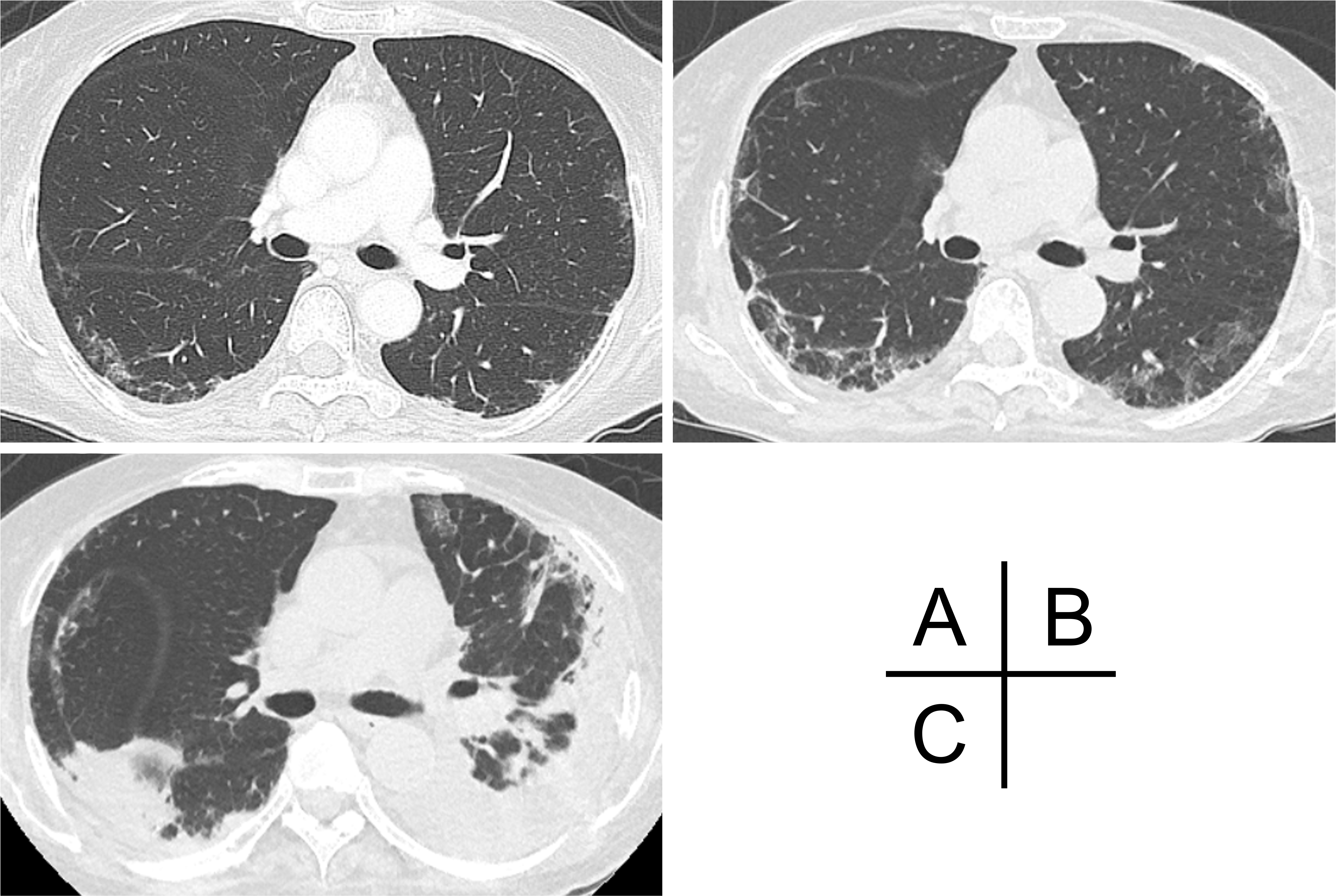
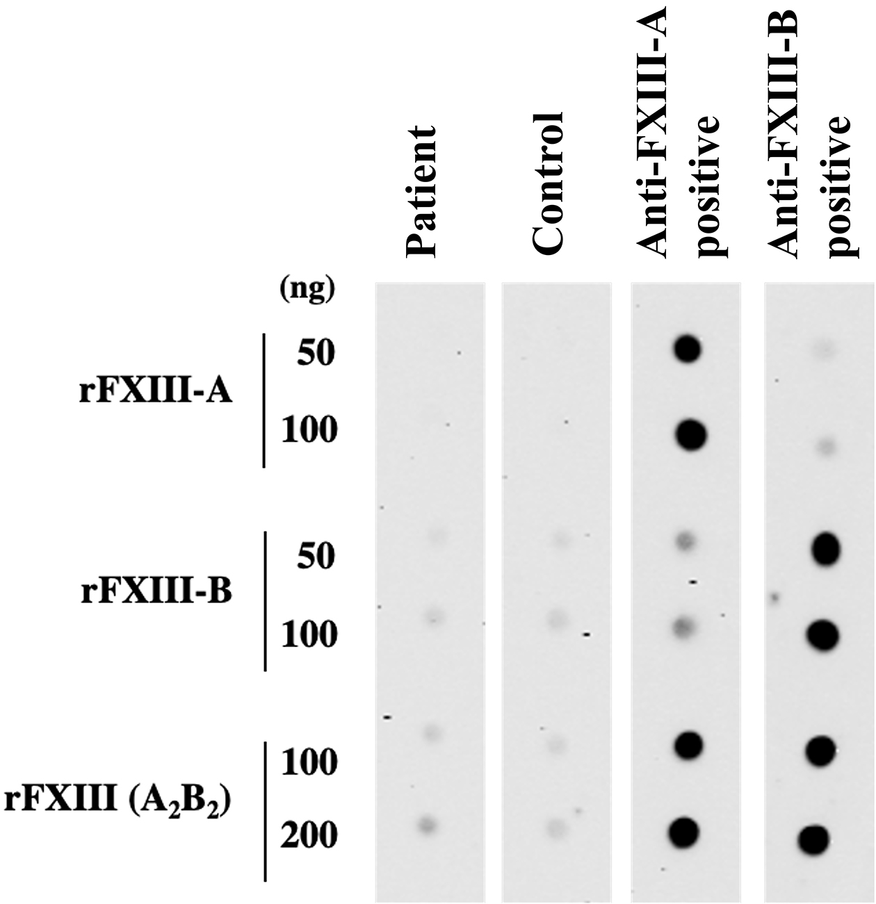
Figure 4. Immunoblotting test for detecting anti-factor XIII (FXIII) autoantibodies. Nitrocellulose membrane-bounded with recombinant FXIII-A subunit (rFXIII-A), recombinant FXIII-B subunit (rFXIII-B) proteins, and their complexes (rFXIIIA2B2) at the indicated amounts was incubated with diluted plasma obtained from healthy control, our patient, acquired FXIII deficiency patient with anti-FXIII-A autoantibody or anti-FXIII-B autoantibody. Immunoglobulin bound to rFXIII was then detected using a peroxidase-labelled anti-human immunoglobulin antibody.
From: Spontaneous Intramuscular Hemorrhage in Anti-melanoma Differentiation-associated Gene 5 Antibody-positive Dermatomyositis