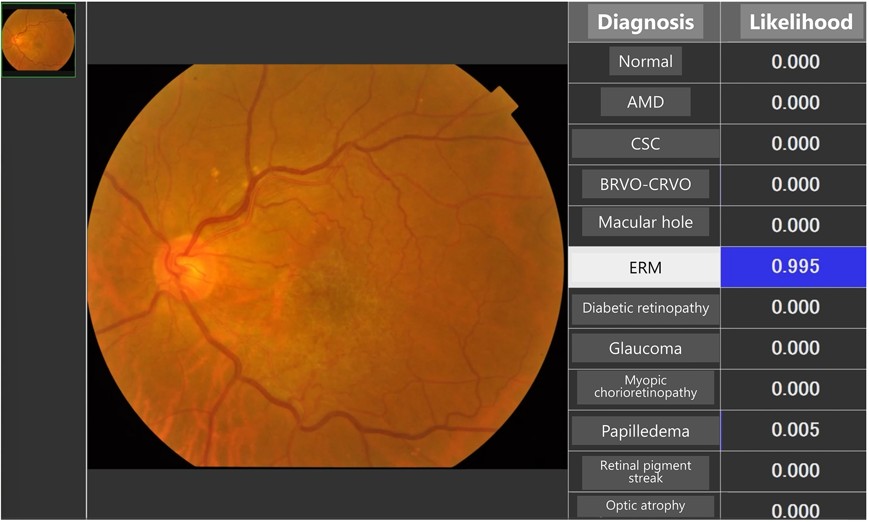
Figure 2. AI application to assist in the differentiation of nine anterior segment conditions. The condition with the highest likelihood is highlighted (8). In cases of infectious keratitis, the most likely pathogen is highlighted (10).
From: Artificial Intelligence Applications in Ophthalmology
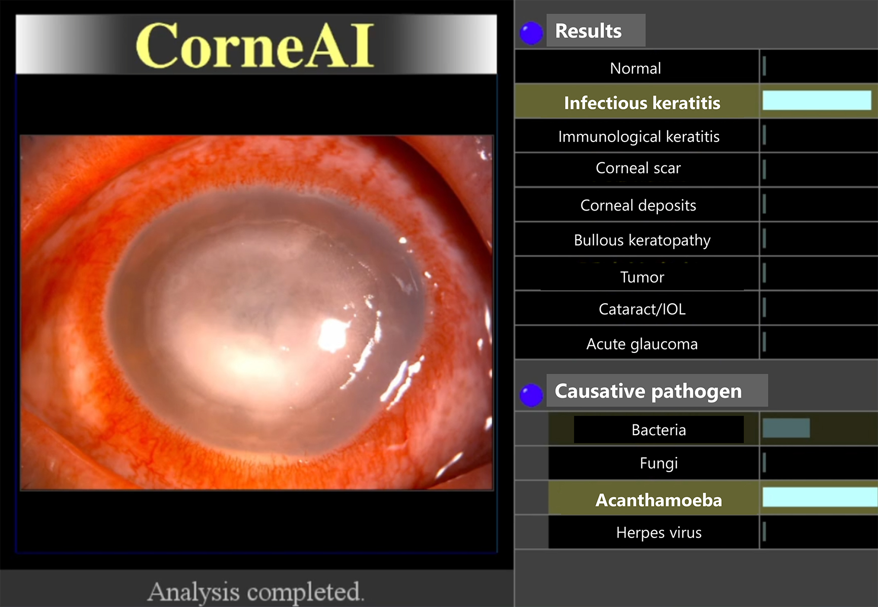
Figure 5. Intraocular lens packages. A mobile device camera is used to capture the image to recognize the parameters shown on the packages of planned, reserved, and backup lenses. The AI system verifies the accuracy of these parameters before surgery (38).
From: Artificial Intelligence Applications in Ophthalmology
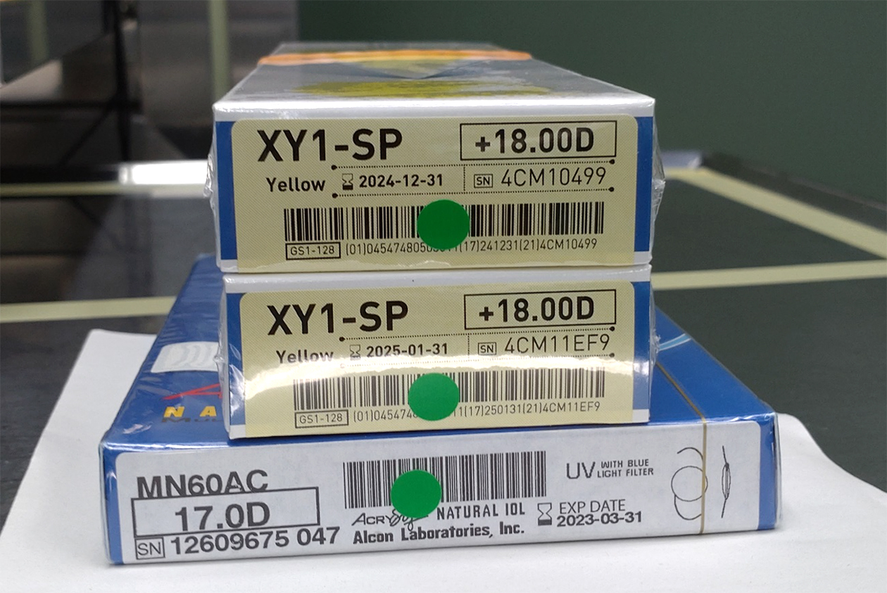
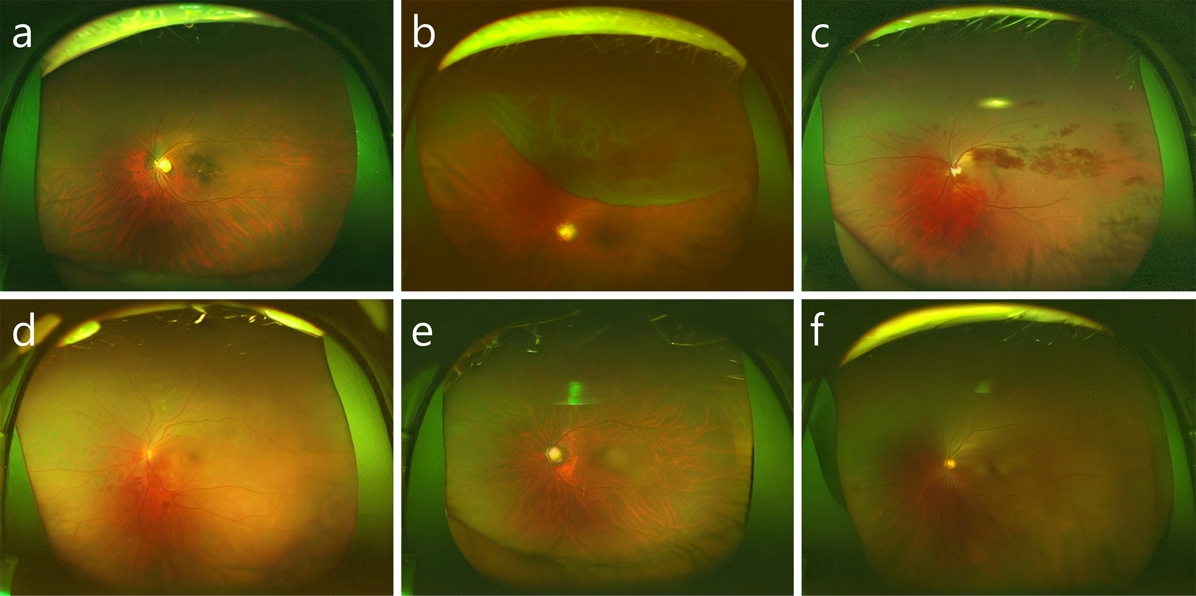
Figure 6. Retinal images were artificially generated using a stable diffusion model. (a) Age-related macular degeneration, (b) retinal detachment, (c) retinal vein occlusion, (d) diabetic retinopathy, (e) glaucoma, and (f) normal fundus.
From: Artificial Intelligence Applications in Ophthalmology