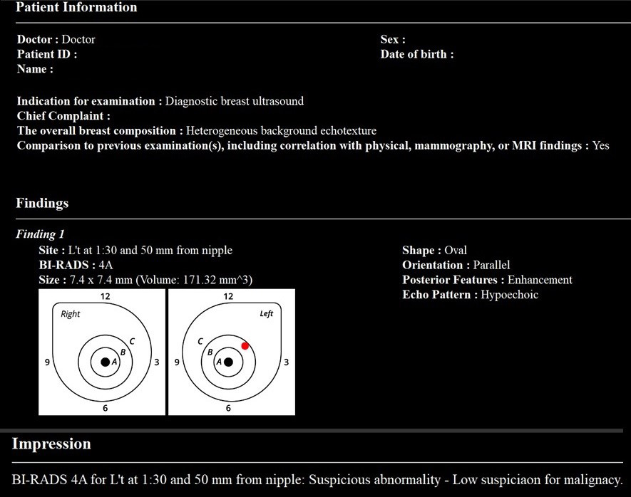
Figure 1. Diagram Showing the Relationships Among AI, ML, and DL
Artificial intelligence (AI) is a broad concept that encompasses machine learning (ML), and within ML, there is deep learning (DL). Deep learning has several models, including convolutional neural networks (CNNs) and large language models (LLMs).
From: The Future of Breast Cancer Diagnosis in Japan with AI and Ultrasonography
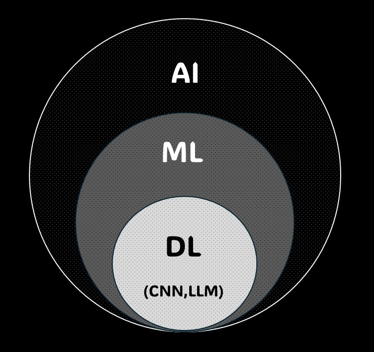
Figure 2. Annual Number of Publications on AI in Breast Ultrasound
This figure shows the annual number of publications on Artificial intelligence (AI) in breast ultrasound from 2014 to 2023. The data, extracted from PubMed using a specific query, indicates a steady number of publications from 2014 to 2016, followed by a significant increase starting in 2017. Publications exceeded 100 annually since 2018, surpassing 300 in 2020, and reaching 699 in 2023.
From: The Future of Breast Cancer Diagnosis in Japan with AI and Ultrasonography
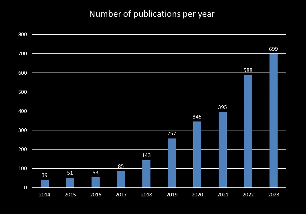
Figure 3. Approaches to CAD-Assisted Image Reading
Three artificial intelligence (AI) reading approaches exist: second-reader, concurrent-reader, and first-reader. The second-reader method involves initial radiologist reading followed by AI reanalysis. The concurrent-reader method uses real-time AI analysis during reading. The first-reader method has AI analyze images first, then the radiologist reviews AI-identified candidates.
From: The Future of Breast Cancer Diagnosis in Japan with AI and Ultrasonography
Figure 4. Detection of Breast Lesions Using CADe in Ultrasound
In November 2020, a breast cancer detection support program was approved for the ultrasound modality in Japan for the first time. Artificial intelligence (AI) has two functions: it performs shape analysis during the examination, displaying suspected breast cancer areas as regions of interest on the image screen. The probe position is also shown on a route map screen.
From: The Future of Breast Cancer Diagnosis in Japan with AI and Ultrasonography
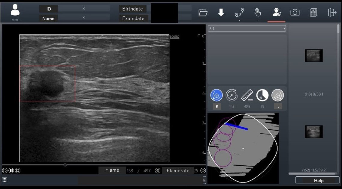
Figure 5. AI Analysis of Mammography Images
A mass is observed in the lower region of the left mediolateral oblique view (→). Artificial intelligence (AI) quantifies the probability of malignancy and highlights areas of interest on a heat map when mammography images are uploaded. This case was diagnosed as left breast cancer. The AI correctly identified it as breast cancer.
From: The Future of Breast Cancer Diagnosis in Japan with AI and Ultrasonography
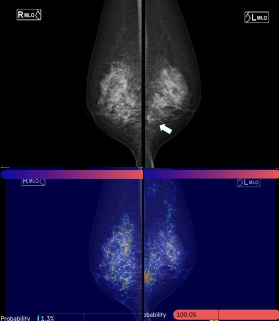
Figure 6. Lesion Identification and Evaluation with an AI System for ABUS
An artificial intelligence (AI) system for Automated Breast Ultrasound (ABUS) can identify lesion candidates and provide precise locations and sizes of lesions. Additionally, it evaluates the shapes and findings of lesions based on BI-RADS (Breast Imaging Reporting and Data System), aiding in diagnosing BI-RADS categories. This figure illustrates how the system has the potential to enhance diagnostic accuracy and efficiency, complementing radiological reading.
From: The Future of Breast Cancer Diagnosis in Japan with AI and Ultrasonography
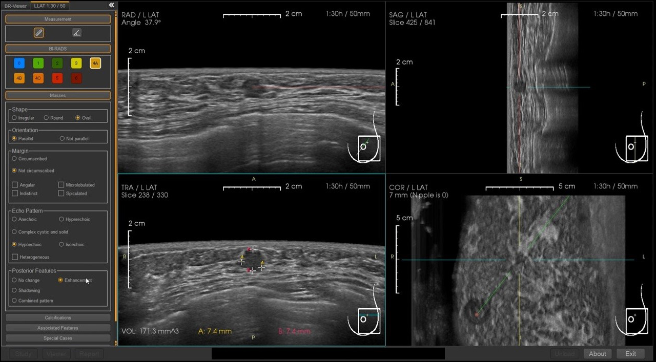
Figure 7. Automatic Selection of Key Images and Report Generation with an AI System for ABUS
An artificial intelligence system for Automated Breast Ultrasound (ABUS) features automatic selection of key images and generation of reading reports, which can significantly reduce the time required for reading and report preparation. This figure demonstrates the potential capability of the system to streamline the diagnostic process, making it more efficient for radiological reading.
From: The Future of Breast Cancer Diagnosis in Japan with AI and Ultrasonography