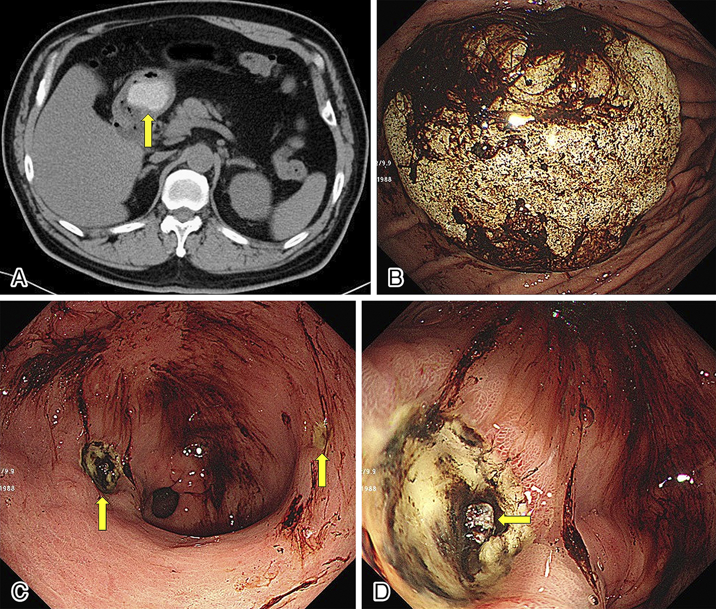
Figure 1. (A) Abdominal computed tomography scan showing a high-density mass (arrow) in the stomach. (B) Esophagogastroduodenoscopy (EGD) showing a whitish solid structure with hematin in the gastric body. (C) EGD showing two gastric ulcers (arrows) and hematin in the antrum. (D) One of the gastric ulcers had a visible vessel (arrow), and hemostatic clips were applied.
From: Hemorrhagic Gastric Ulcer due to the Stagnation of Rice Cake