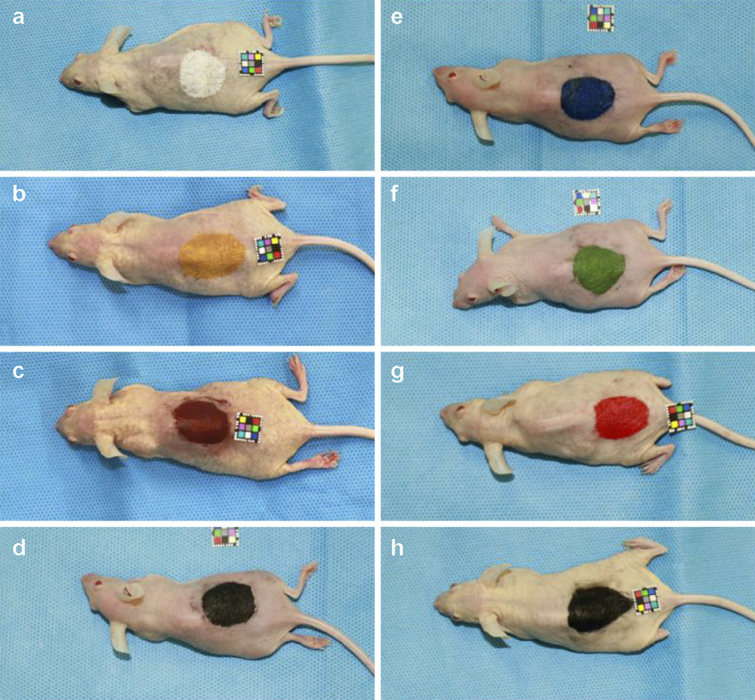
Figure 1. Mice tattooed with pigments containing metal components. A 25-mm circular tattoo was applied to the back of hairless Hos:HR-1 mice. The utilized colors were: (a) titanium dioxide; (b) iron oxide yellow; (c) iron oxide red; (d) iron oxide black; (e) ultramarine blue #29; (f) chromium oxide green #7; (g) D&C red #7; and (h) carbon black.
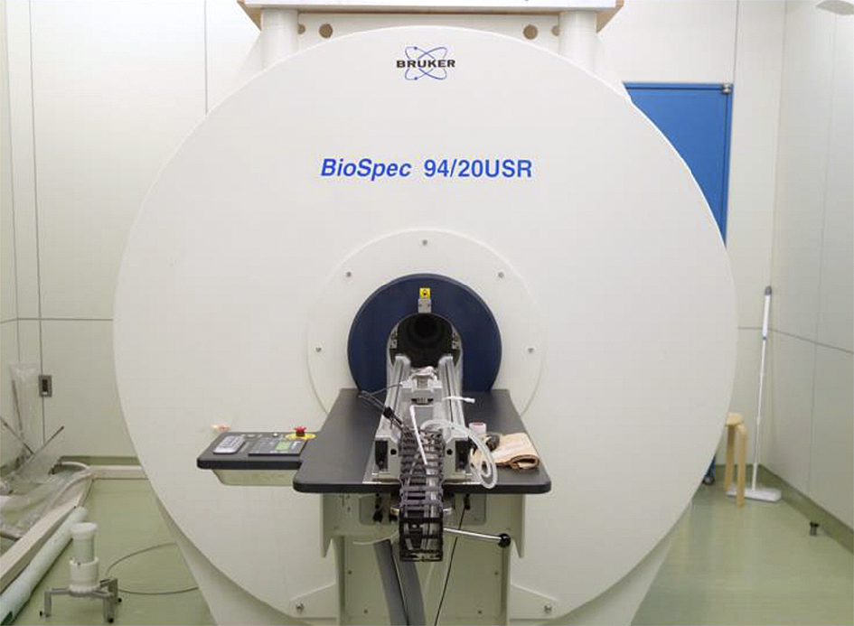
Figure 2. An ultra-high magnetic field magnetic resonance scanner (9.4-T) manufactured by Bruker (BioSpec 94/20 USR) was used for magnetic resonance imaging. The sequence was as follows: repetition time 3,500 msec; Echo time 11.0 msec; Flip angle, 180°.
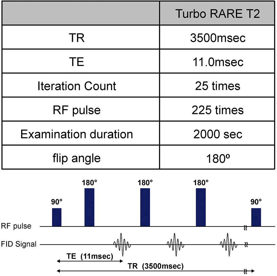
Figure 3. Sequence of the magnetic resonance imaging (MRI). The spin echo method used to obtain a radiofrequency (RF) pulse echo was the Turbo RARE T2 sequence.
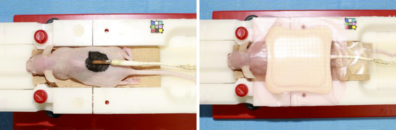
Figure 4. Surface temperature measurement during the magnetic resonance imaging procedure. The body temperature probe was applied to the tattooed area, which was insulated on one side using polyurethane foam. The animal was placed in the gantry of the MRI scanner (Figure 2) for more than 30 min; when the temperature measurement plateaued, the MRI began.
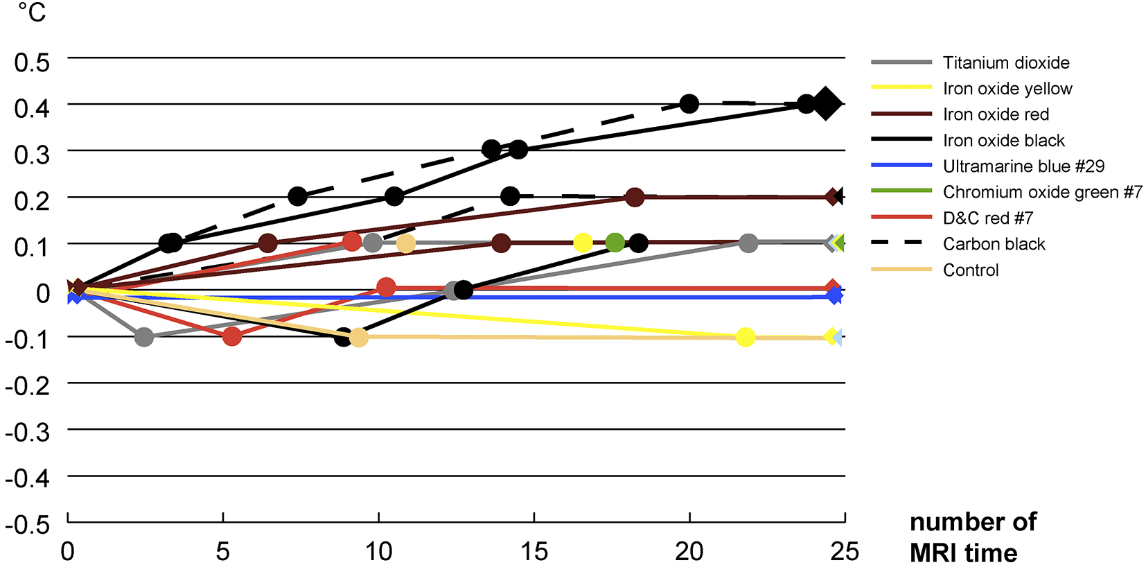
Figure 5. Temperature changes during the magnetic resonance imaging procedure. Temperature changes on the biological monitor of the MRI operation room were recorded continuously.
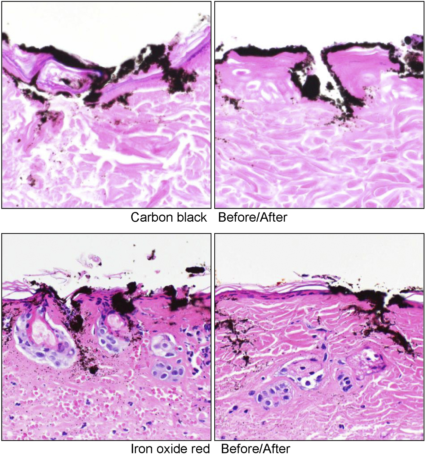
Figure 6. Tissue specimens before and after the magnetic resonance imaging examination. The pigments were found not only in the dermis, but also in the epidermis. It was confirmed that there was erosion of the epidermis. Neutrophil infiltration and the denaturation of collagen fibers were observed in the dermal layer.