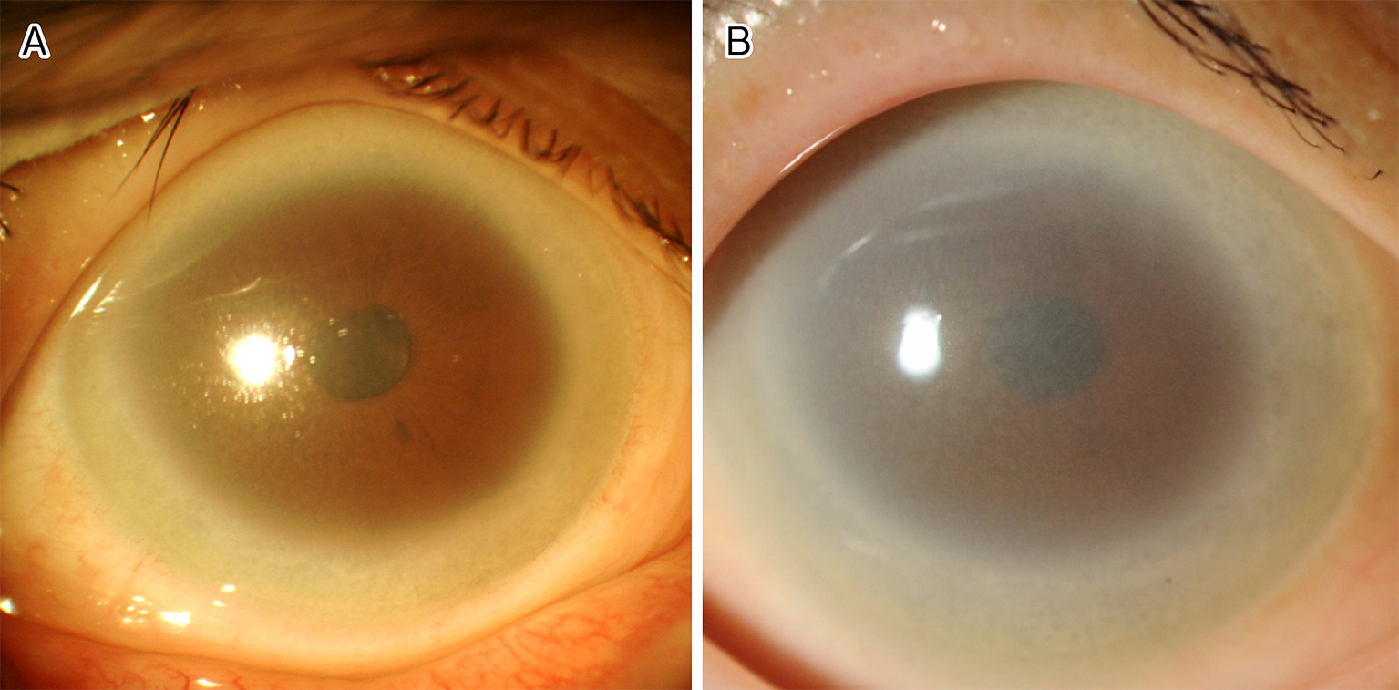
Figure 1. (A) A slit-lamp photograph showing the cornea with marked corneal opacification in the peripheral area. (B) A slit-lamp photograph showing the cornea of the same patient after 15 years of observation. The corneal opacity has become denser and has progressed to the central area. It was difficult to observe the fundus.
From: Bilateral Corneal Opacity of Fish-eye Disease