Figure 2. Sagittal Gd-enhanced T1-weighted MRI. Spinal epidural abscess is noted that a capsule-like rim whose edges are strongly enhanced and the content of abscess shows low intensity in the cervical (A) and thoracic (B) spine. The retro-pharyngeal abscess is also noted as enhanced mass (A). Arrowheads indicate the abscess.
From: Spinal Epidural Abscess: A Review Highlighting Early Diagnosis and Management
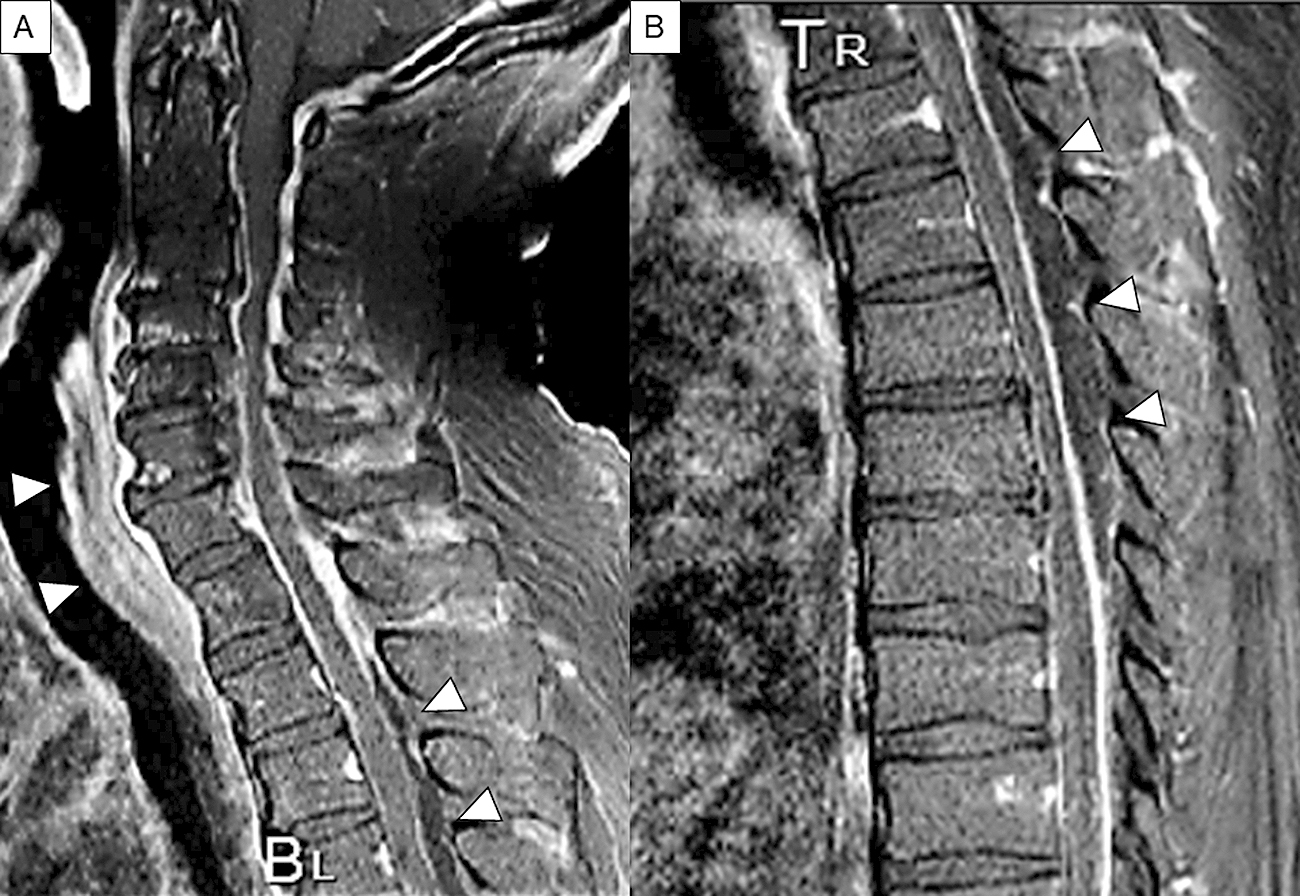
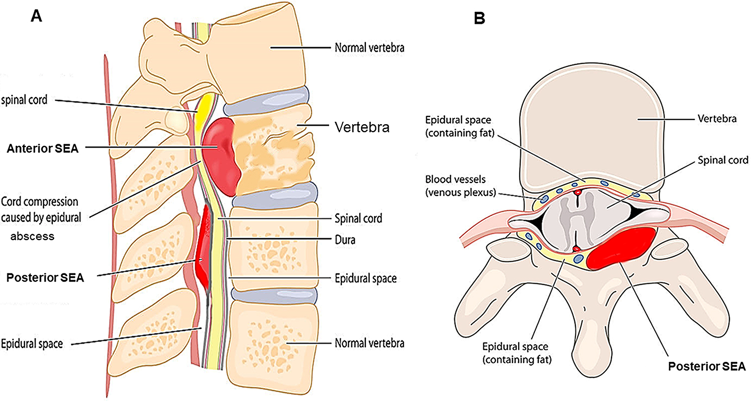
Figure 3. Sagittal and axial views of T1-weighted, T2-weighted, and Gd-enhanced T1-weighted MRI. Extensive epidural phlegmon is noted throughout the lumbar spine in the sagittal view; T1-weighted images show low signal intensity (A), T2-weighted images show high signal intensity (B), and Gd-enhanced MRI shows that a linear structure appears to be a thickened dural surface on the ventral side of the lamina. Acknowledge the occupying lesions inside and capsule-like rims whose edges are strongly enhanced. (C). The axial view at the level of L4 confirms that the dural tube is compressed by posterior SEA in each image (D–E). The enhancement is also observed in the L5 vertebral body and the paraspinal muscles, representing spondylitis and spread of inflammation into the surrounding muscles (C, F). Arrowheads indicate SEA.
From: Spinal Epidural Abscess: A Review Highlighting Early Diagnosis and Management
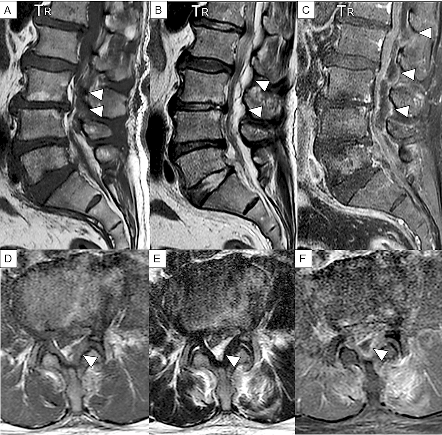
Figure 4. Sagittal and axial T2-weighted MRI in case 1.
Extensive epidural phlegmon is noted throughout the lumbar spine in the sagittal view (A) and in the axial view (B). Content of abscess shows heterogeneous high intensity, accompanied by low intensity of capsule-like rim, which indicate granulation tissue (A). The axial view confirms that the dural tube is compressed posteriorly at the level of L3 (B). Arrowheads indicate SEA.
From: Spinal Epidural Abscess: A Review Highlighting Early Diagnosis and Management
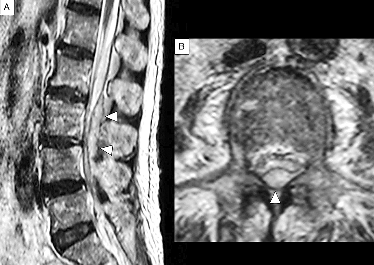
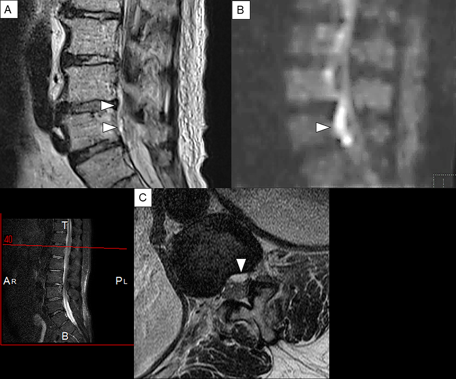
Figure 5. Sagittal T2-weighted and Gd-enhanced MRI of the lumbar spine in case2.
(A): Sagittal view of T2-weighted MRI.(B): Sagittal view of diffusion-weighted image. (C): Axial view of T2-weighted MRI. A linear lesion that shows high intensity and appears to be an epidural abscess that is found on the dorsal side of the vertebral body (A). The same lesion shows a strong high signal area in the diffusion-weighted image (B). This finding is considered to reflect the decrease in diffusion of water molecules in the abscess lumen and is characteristic findings of abscess. The axial view confirms anterior SEA that shows high signal intensity on the dorsal side of the vertebral body at the level of L1 (C). Arrowheads indicate SEA.
From: Spinal Epidural Abscess: A Review Highlighting Early Diagnosis and Management