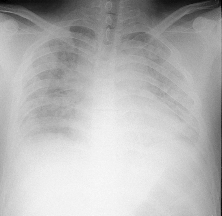
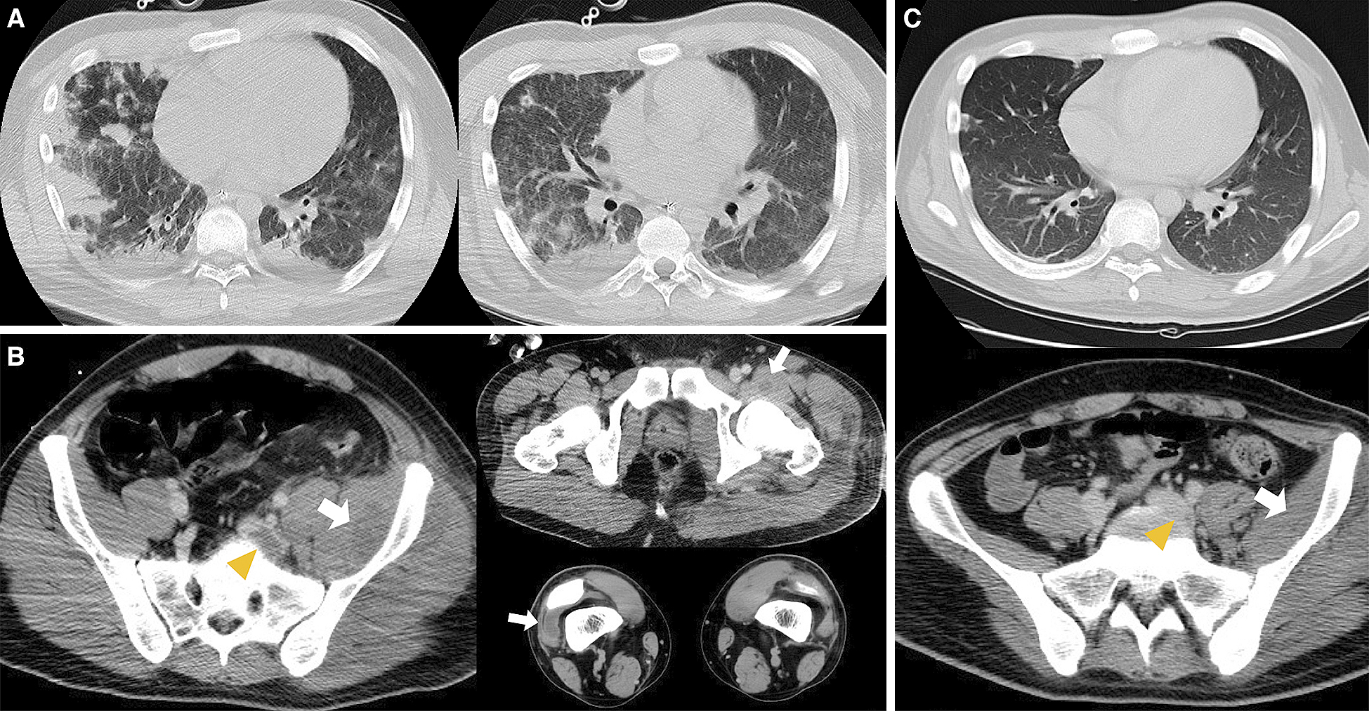
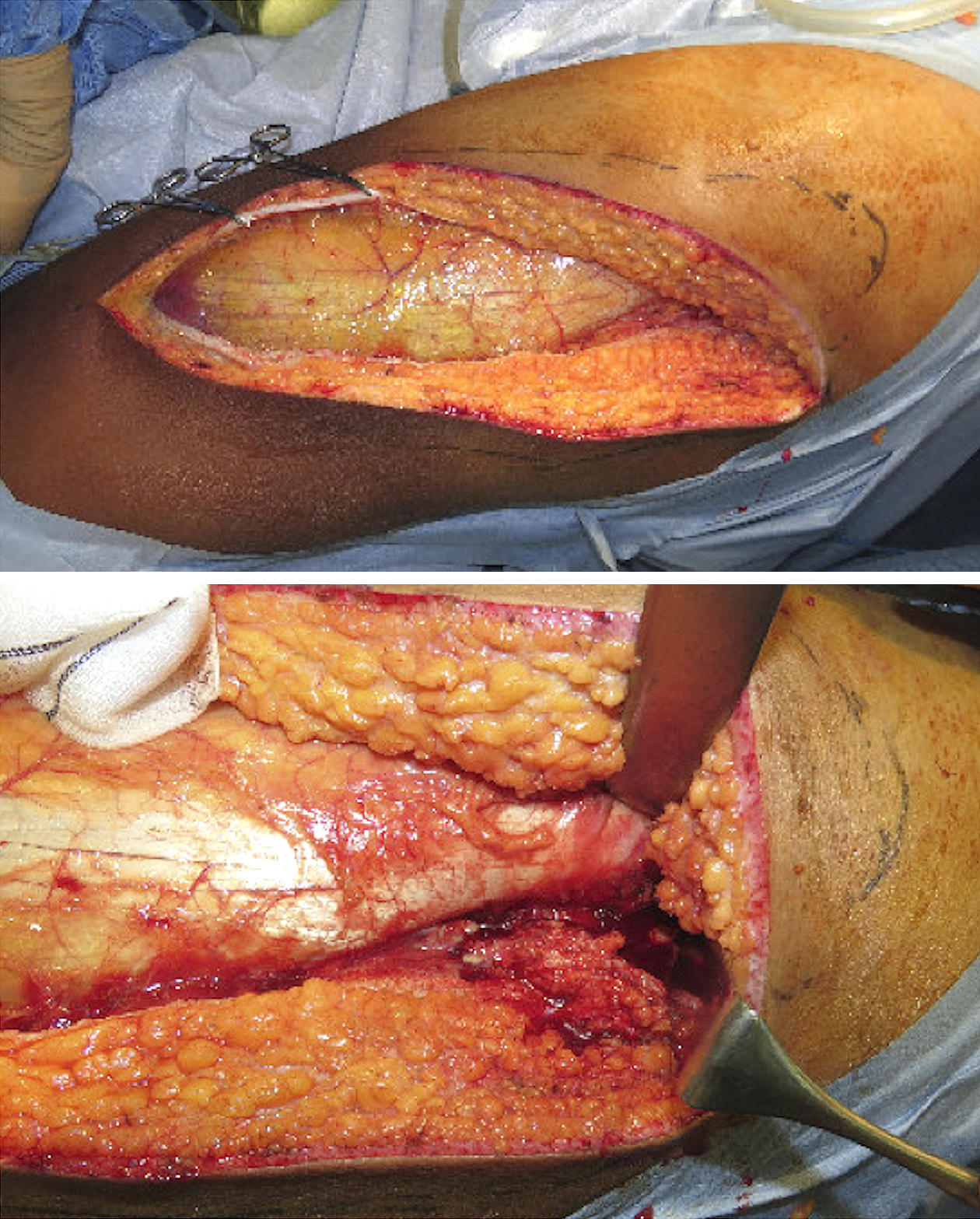
Figure 2. A) Contrast-enhanced computed tomography of the chest revealed septic pulmonary emboli. B) Contrast-enhanced computed tomography revealed left iliofemoral thrombosis (arrow head) and skin and soft tissue infection of the hip and left thigh with abscesses (arrow). C) CT after 1 month revealed the resolution of the iliofemoral deep venous thrombosis (arrow head).