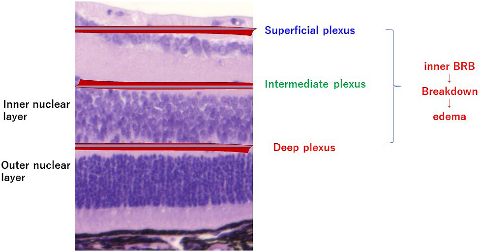
Figure 1. Retinal blood vessels. Retinal capillaries form three major layers. The intermediate capillary plexus is formed between the inner plexiform layer and inner nuclear layer, and the deep capillary plexus is formed in the outer plexiform layer. The retinal capillaries constitute the inner blood retinal barrier (BRB). Breakdown of the inner BRB causes edema.
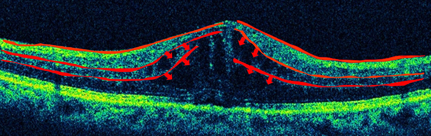
Figure 2. Diabetic macular edema. Edema presumably arises from the capillaries in the intermediate and deep plexuses of the retina, which constitute the inner BRB.
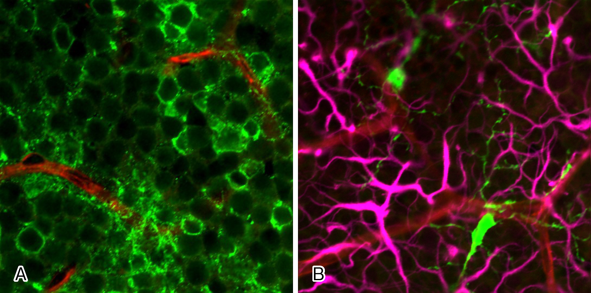
Figure 3. Neurites of retinal amacrine cells, microglia, and capillaries of intermediate plexus of retina. (A) Neurites (green) of numerous amacrine cells (green) are in contact with the capillaries of the intermediate plexus (red). (B) Retinal microglia (green) are in contact with the capillaries of the intermediate plexus (red) and with neurites of amacrine cells (pink).
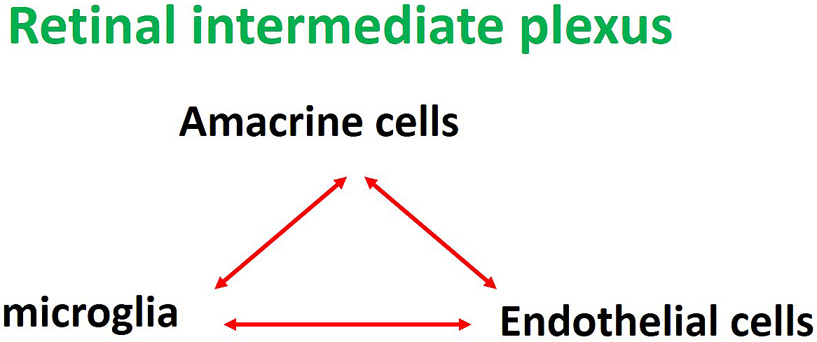
Figure 4. Neurovascular units in the intermediate plexus of retina. Retinal amacrine cells, microglia, and vascular endothelial cells constitute the neurovascular units in the intermediate plexus of the retina.
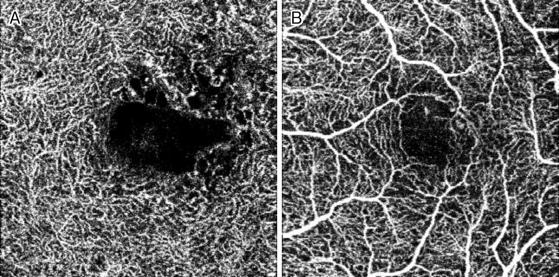
Figure 5. OCT angiography images (Topcon, DRI OCT Triton SSOCT AngioTM, 3×3 mm) of diabetic macular edema. The retinal capillaries in the intermediate or deep plexuses (A), but not superficial plexuses (B), are partially lost.
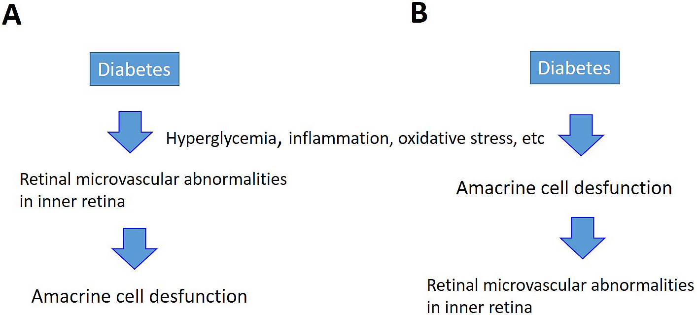
Figure 6. Diabetes-induced damages to blood vessels and amacrine cells in the inner layers of retina. Two pathways are speculated. (A) Hyperglycemia, inflammation, and oxidative stress directly damage capillaries in the retina. (B) Primary damage to neurons that control capillaries in the retina results in secondary damage to retinal vascular endothelial cells.