Figure 1. Immune response in ischemia-reperfusion injury kidney.
Both innate and adoptive immune systems are involved in ischemia-reperfusion injury model.
Kidney injury occurred according to the timeline. Immune system activated in post-ischemic kidneys by resident dendritic cells and macrophages. Disruption of endothelial cells causes an influx of immune cells to damaged kidneys.
Abbreviations: NK cell; natural killer cell, Treg cell; regulatory T cell.
From: Neuroimmune Communication in the Kidney
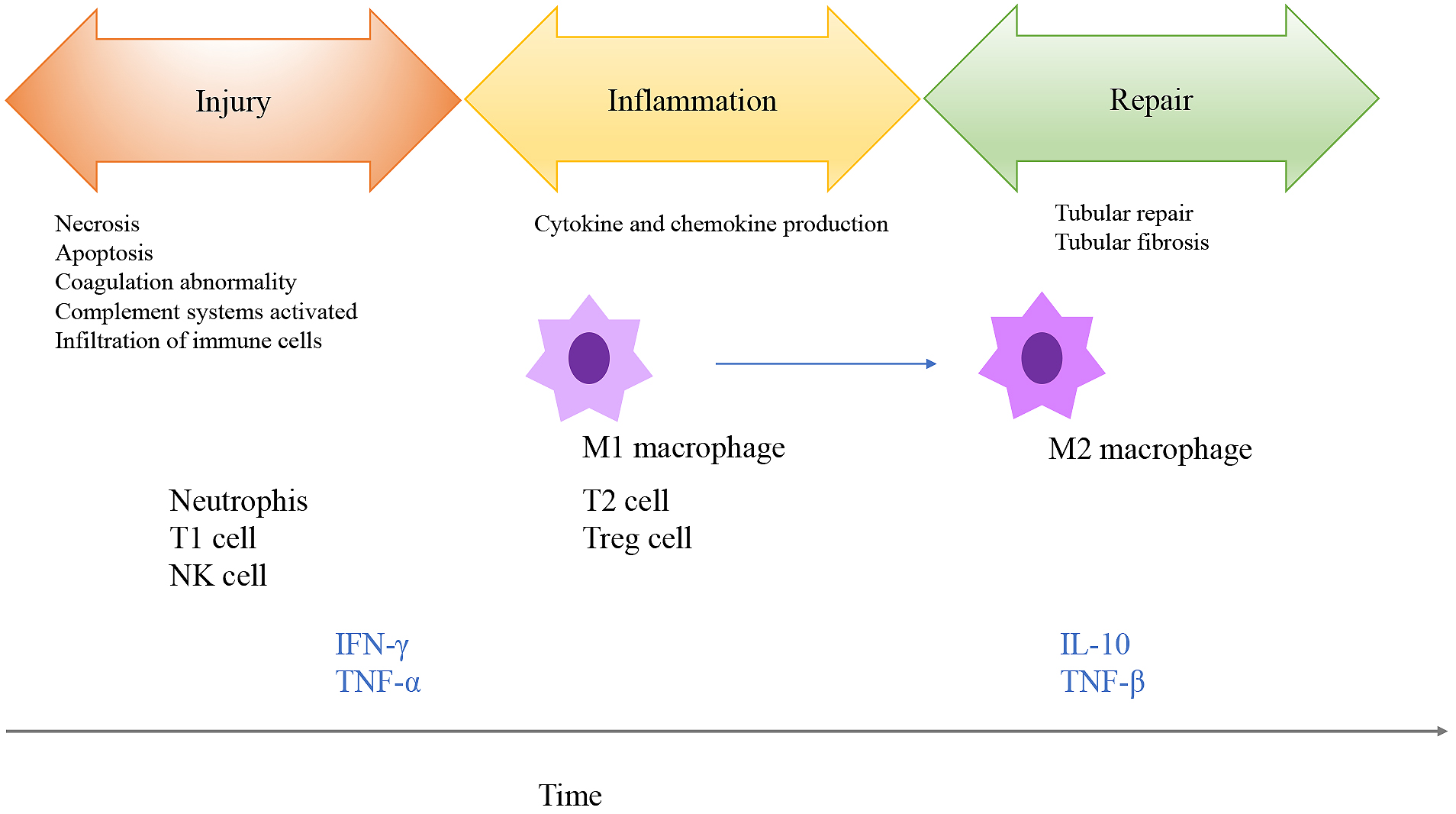
Figure 2. Inflammatory pathway.
There are several pathways involved in inflammatory response. HPA axis and sympathetic/parasympathetic nerves are linked.
Abbreviations: CNS; central nervous system, HPA axis; hypothalamic-pituitary-adrenal axis, ACTH; adrenocorticotrophic hormone, ACh; acetylcholine, NA; noradrenaline.
From: Neuroimmune Communication in the Kidney
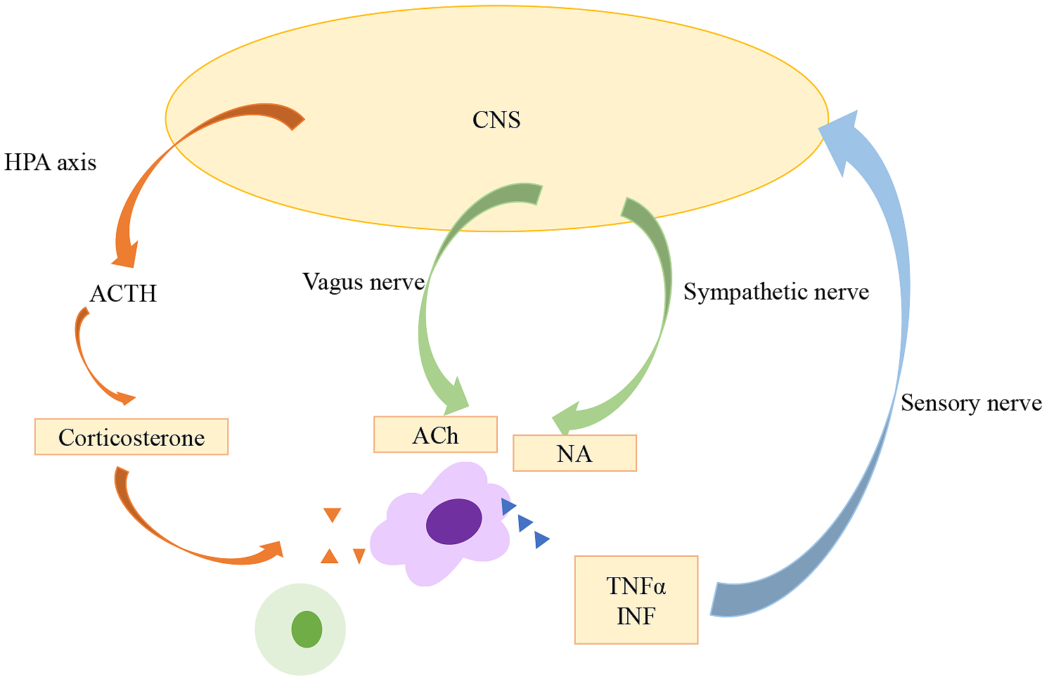
Figure 3. Overview of cholinergic anti-inflammatory pathway (CAP).
Once local inflammation produces proinflammatory cytokines, afferent vagus nerves detect these inflammatory signals and transmit them to NTS to DMV in medulla oblongata. These signals are transferred to the spleen via efferent vagus nerve (unknown). Noradrenaline released from the terminal of splenic nerve, binds to β2 adrenergic receptors in the CD4 positive ChAT T cells. Acetylcholine released from these cells binds to α7 nicotinic receptors in macrophages, resulting in decrease in inflammatory responses.
Abbreviations: CNS; central nervous system, NTS; nucleus tractus solitarius, DMV: dorsal motor nucleus of the vagus, NA; noradrenaline, ACh; acetylcholine, ChAT; choline acetyltransferase, PAMPs; pathogen-associated molecular patterns, DAMPs; damage-associated molecular patterns.
From: Neuroimmune Communication in the Kidney
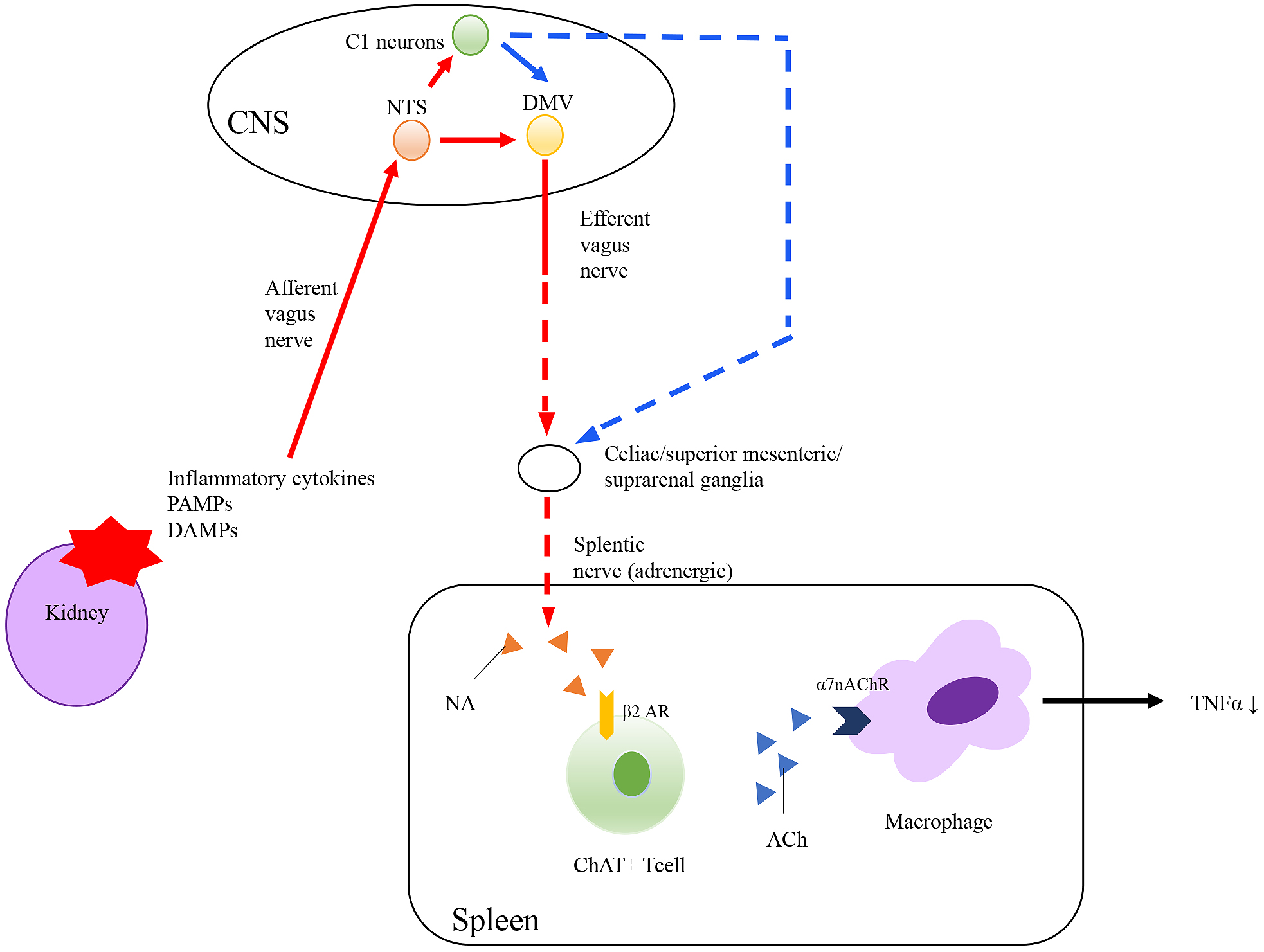
Figure 4. Innervation in Kidney.
Sympathetic nerves and sensory nerves innervate in kidney. Innervation of parasympathetic nerves in kidney is controversial.
From: Neuroimmune Communication in the Kidney
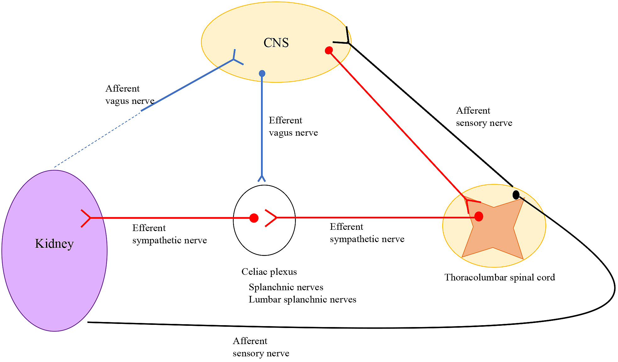
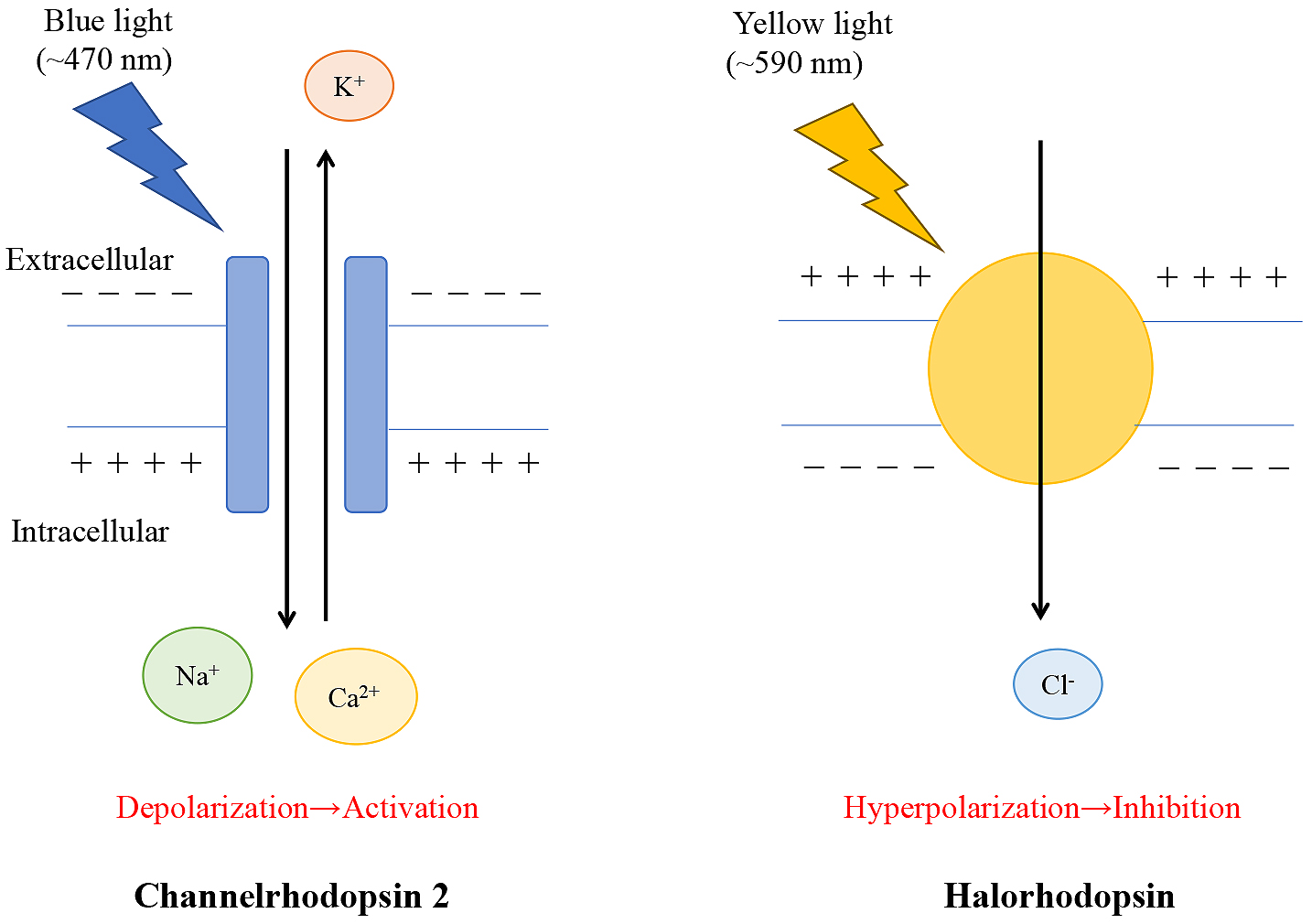
Figure 5. Optogenetics.
Channelrhodopsin 2 or halorhodopsin are introduced genetically into specific cells or neurons for optogenetic stimulation. Blue light opens to channelrhodopsin 2. Then, cations flow into the specific neurons through the membrane. This evokes cell depolarization and eventually leads to neuronal firing. Yellow light opens to halorhodopsin, and one chloride ion enters the target cell through the membrane, hyperpolarizing the specific neuron.
Selective cation channel (a chloride pump), resulting in excitation (inhibition) of the neurons.
From: Neuroimmune Communication in the Kidney