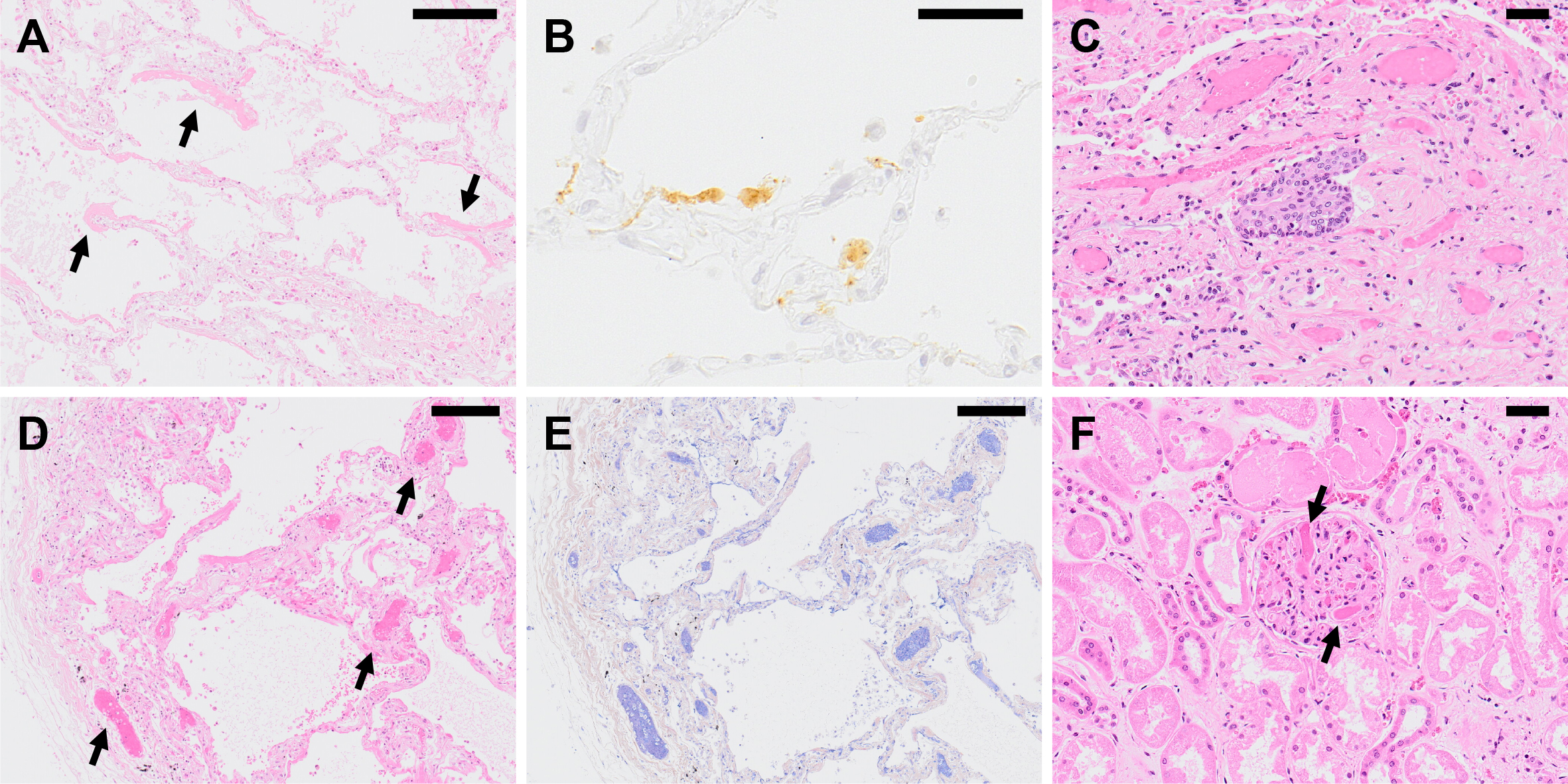
Figure 1. Histopathological findings of COVID-19 pneumonia. (A) A lung specimen of COVID-19 pneumonia obtained from an autopsied case showing exudative phase of DAD with formation of hyaline membrane (arrow) along alveolar septa. Hematoxylin & eosin (H&E) staining. Scale bar indicates 200 μm. (B) Detection of SARS-CoV-2 antigen in COVID-19 pneumonia by immunohistochemistry using a rabbit polyclonal antibody against SARS-CoV-2 antigens (65). Positive signals (brown) are observed on alveolar epithelial cell and alveolar macrophages. Scale bar indicates 50 μm. Panels A and B are from the same case at the same time. (C) A lung specimen of COVID-19 pneumonia showing organizing phase of DAD with squamous cell metaplasia. H&E staining. Scale bar indicates 50 μm. (D) Exudative phase of DAD accompanied with prominent microvascular thrombosis (arrow) observed in COVID-19 pneumonia. H&E staining. Scale bar indicates 200 μm. (E) Microthrombi (blue) are highlighted by phosphotungstic acid hematoxylin (PTAH) staining on the same specimen as panel D. Scale bar indicates 200 μm. (F) Microthrombi formation (arrow) in a glomerulus of the kidney from a COVID-19 autopsy case. H&E staining. Scale bar indicates 50 μm.
From: Insights into Pathology and Pathogenesis of Coronavirus Disease 2019 from a Histopathological and Immunological Perspective