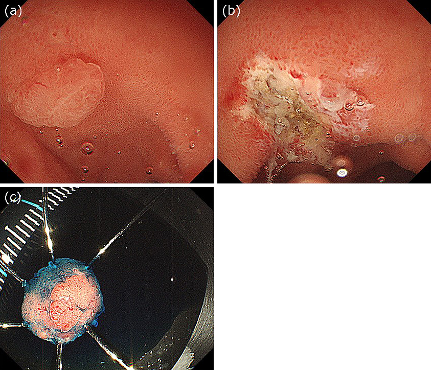
Figure 1. (a) Endoscopic image showing two adenocarcinomas in the duodenum. One lesion is on the superior duodenal angle (SDA), and the other is on the posterior wall of the duodenal bulb. (b) Narrow-band imaging view of the tumor on the SDA. (c) Narrow-band imaging view of the tumor on the duodenal bulb.
Figure 2. (a) Gel was injected into the duodenal lumen through the accessory channel, and the tumor was immersed. (b) The tumor was completely removed. (c) The resected specimen.