Figure 1. (Case 1) Contrast-enhanced computed tomography showed a heterogeneous tumor in the second portion of the duodenum (arrow).

From: Wedge Resection for Duodenal Gastrointestinal Stromal Tumors: Surgical Management and the Clinicopathological Outcome

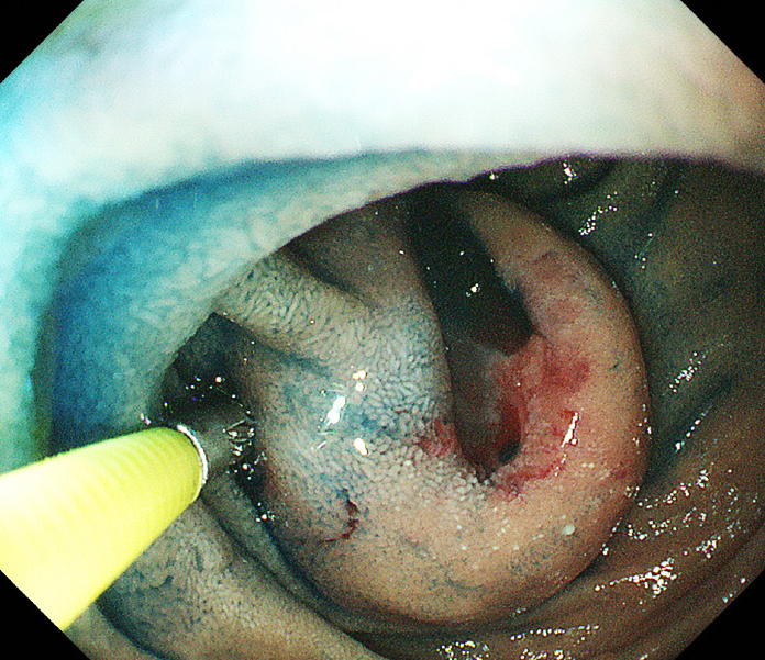
Figure 2. (Case 1) Esophagogastroduodenoscopy showed a tumor with ulceration in the second portion of the duodenum, which was definitely diagnosed by a biopsy.

From: Wedge Resection for Duodenal Gastrointestinal Stromal Tumors: Surgical Management and the Clinicopathological Outcome

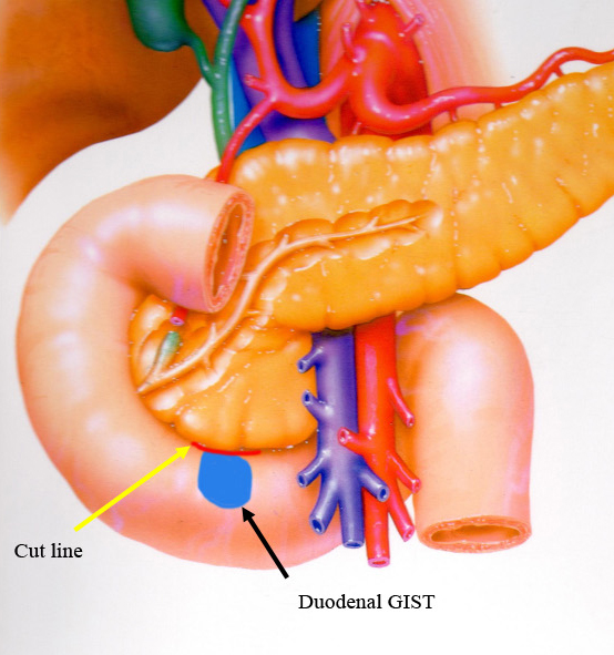
Figure 3. Wedge resection of duodenal gastrointestinal stromal tumors (dGISTs). dGIST of on the side of the pancreas (black arrow). Cut line between the duodenum and the pancreas (yellow arrow).

From: Wedge Resection for Duodenal Gastrointestinal Stromal Tumors: Surgical Management and the Clinicopathological Outcome

PAGE TOP