Figure 1. Computed tomography images of the chest and head. A) Chest computed tomography (CT) image taken on admission shows slightly left and bottom dominant ground-glass appearance and pleural fluid. B) Head CT images taken on the same day show no obvious infarction or hemorrhage.
From: Pathologic and Neuropathologic Study of a Case of COVID-19
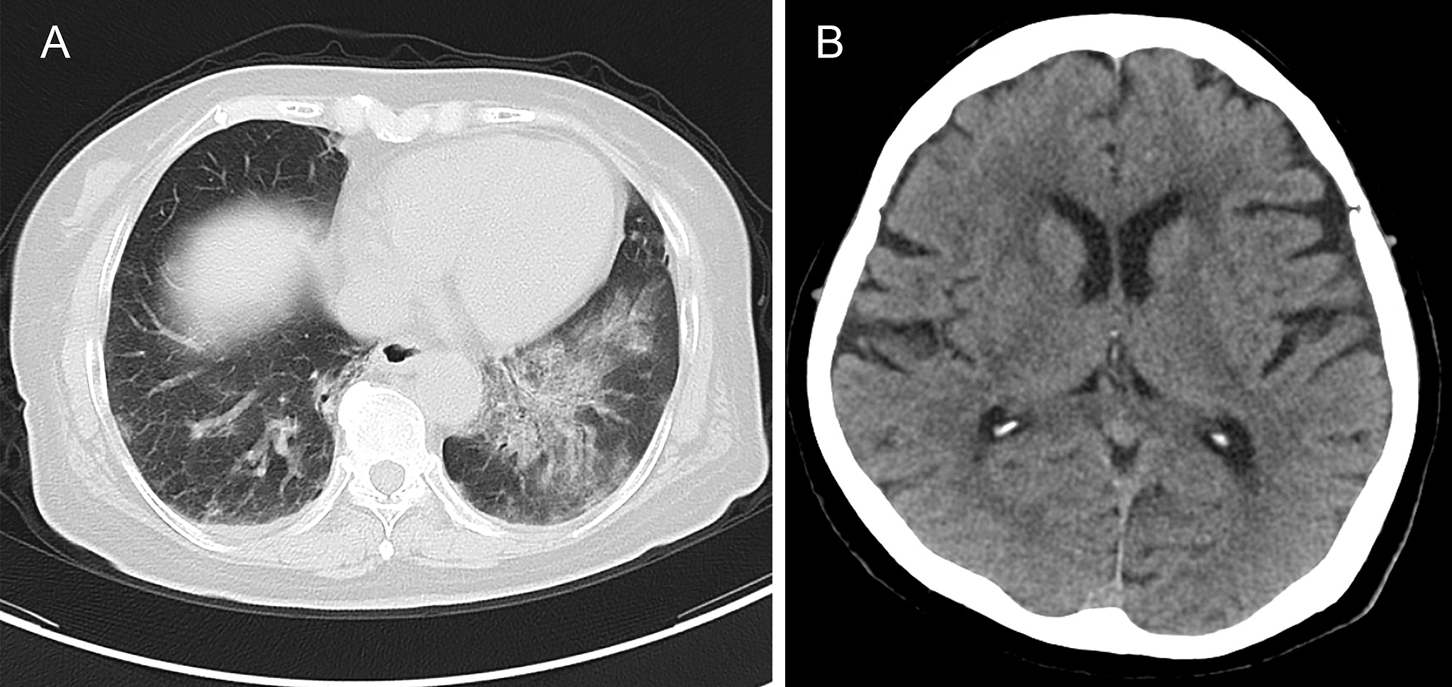
Figure 2. Photomicrographs of the lung and spleen. A) The lung shows diffuse alveolar damage in the exudative to proliferative phases and remodeling of the alveolar wall. B) A small number of fibrin thrombi are present in the small- and medium-sized vessels of the lung. C) The spleen shows acute splenitis. A thrombus adhering to the injured endothelium is present in areas with a high degree of neutrophil infiltration. Scale bar: A: 500 μm; B: 100 μm; C: 50 μm.
From: Pathologic and Neuropathologic Study of a Case of COVID-19
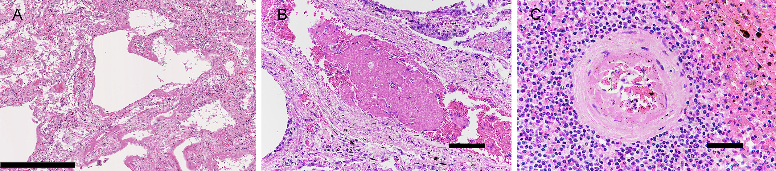
Figure 3. Photomicrographs of the cerebrum. A) An infarct in the white matter of the parietal lobe. B) A small infarction is observed in the thalamus. Inset; close to the lesion, a fibrin thrombus is present in a vessel (arrowhead), as shown in the inset. C) An organized thrombus in the leptomeningeal vessel of the frontal lobe. The thrombi shown in B and C are similar to those shown in Fig. 2B. D, E) Clustered neutrophils in the left parietal cortex and F) internal capsule. Scale bar: A: 1000 μm; B, C, D: 100 μm; B (inset), E, F: 10 μm.
From: Pathologic and Neuropathologic Study of a Case of COVID-19
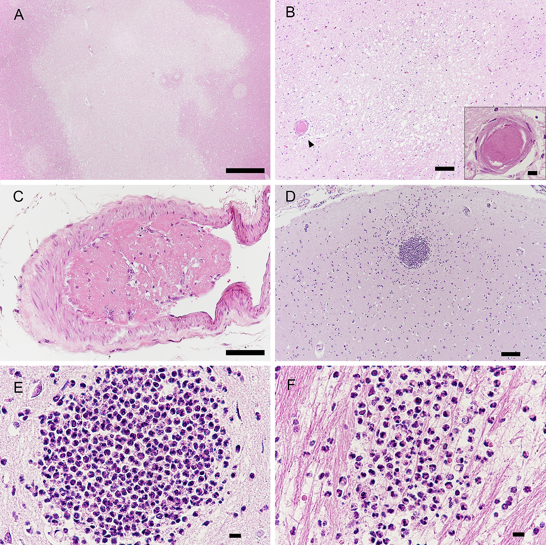
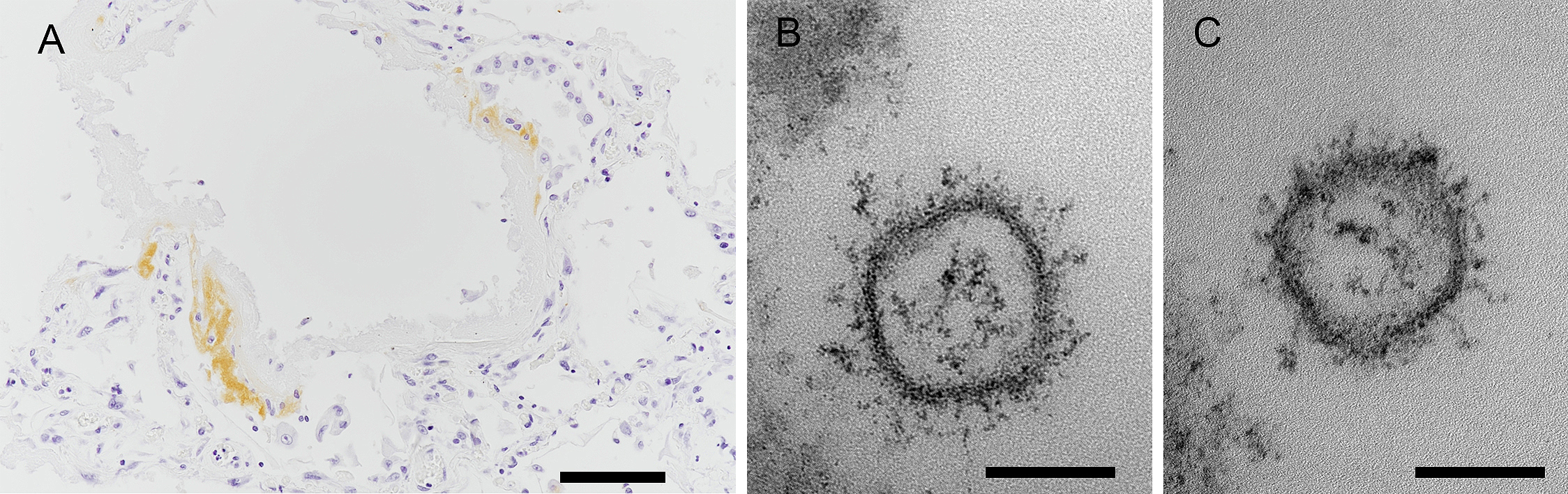
Figure 4. Immunohistochemistry and electron micrograms. A) Immunohistochemistry of the right lung shows that hyaline membranes are positive for the SARS-CoV-2 nucleocapsid. B, C) Small vesicular structures suspected to be SARS-CoV-2 observed by electron microscopy in the extracellular space of the lung. Scale bar: A: 100 μm; B, C: 100 nm.
From: Pathologic and Neuropathologic Study of a Case of COVID-19