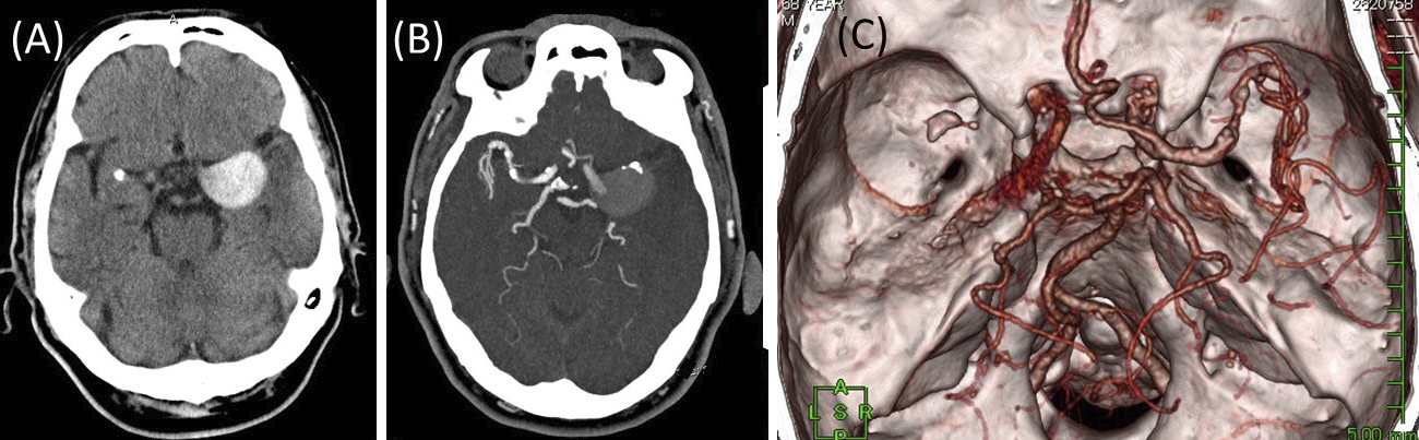
Figure 1. Computed tomography (CT) on admission revealed a high-density, round mass in the left middle cerebral artery (MCA), which was suspected to be an unruptured giant cerebral aneurysm (A). CT angiography revealed MCA occlusion and an aneurysm in the proximal part of the left MCA with poor opacification, suggesting partial thrombosis (B, C).
From: Giant Cerebral Aneurysm Rupture in an Ischemic Stroke