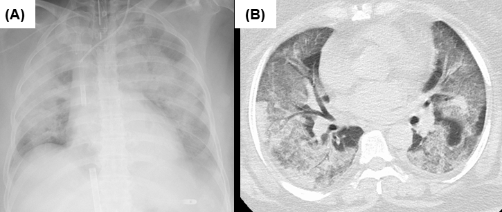
Figure 1. Chest radiograph and computed tomography on admission.
(A) Chest radiograph right after cannulation of ECMO, which showed extensive ground-glass and infiltrative shadows in bilateral lung fields. A drainage cannula was placed in the inferior vena cava and a return cannula was placed in the superior vena cava. (B) Chest computed tomography on admission showed extensive ground-glass shadows in both lungs with partially overlapping infiltrative shadows. Interlobular septal wall thickening and traction bronchiectasis are unremarkable.
From: Alarming of Severe Respiratory Failure Requiring ECMO Caused by the SARS-CoV-2 Omicron Variant