Figure 1. Aβ42 deposition precedes that of Aβ40 in the progression of AD changes in Down syndrome brains.
Frontal cortices from autopsied brains from patients with Down syndrome who died at the ages of 31 (A, B), 44 (C, D), and 57 years (E, F) were immunostained with Aβ42- (A, C, E) and Aβ40-specific monoclonal antibodies.

From: Molecular Pathogenesis and Disease-modifying Therapies of Alzheimer’s Disease and Related Disorders
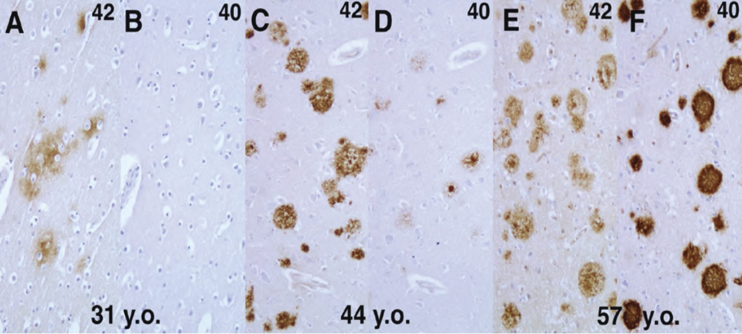
Figure 2. Schematic depiction of the process of γ-secretase complex formation.

From: Molecular Pathogenesis and Disease-modifying Therapies of Alzheimer’s Disease and Related Disorders
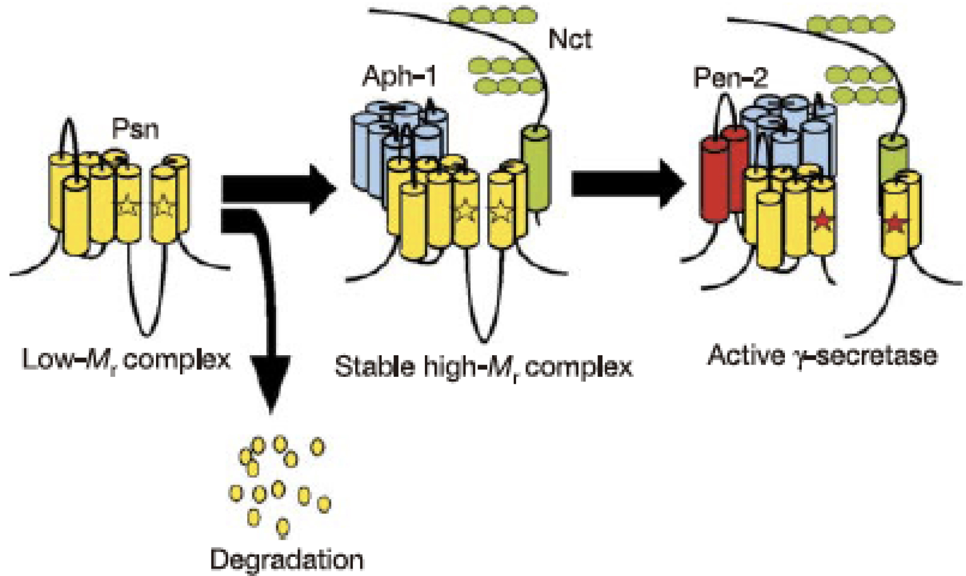
Figure 3. Histopathological and biochemical features of a-synuclein deposited in PD and DLB brains. Immunohistochemistry of cortical-type Lewy bodies (large arrows) and Lewy neurites (small arrows) in DLB cortices (A) and brain stem-type Lewy body in the substantia nigra of PD brain (B) labeled by a phosphoserine 129-specific anti-α-synuclein antibody. C: Immunoblot analysis of sarkosyl-insoluble fractions from control (C) and DLB (D) cortices with a phosphoserine 129-specific anti-α-synuclein antibody (C).

From: Molecular Pathogenesis and Disease-modifying Therapies of Alzheimer’s Disease and Related Disorders
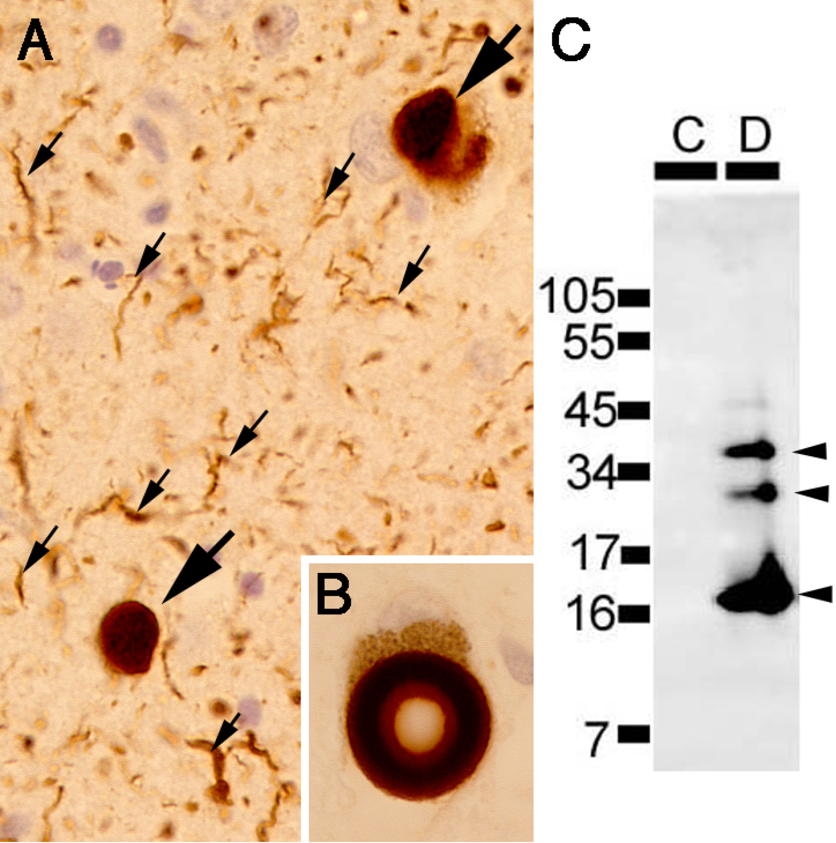
Figure 4. Comparison of cognitive changes (Alzheimer's Disease Assessment Scale-Cognitive Subscale: ADAS-Cog13; upper panel) and clinical progression (Clinical Dementia Rating-sum of boxes: CDR-SB; lower panel) in late MCI due to AD (LMCI) in the J-ADNI (green lines) and the North American ADNI (blue lines); cited from Iwatsubo et al. (23).

From: Molecular Pathogenesis and Disease-modifying Therapies of Alzheimer’s Disease and Related Disorders
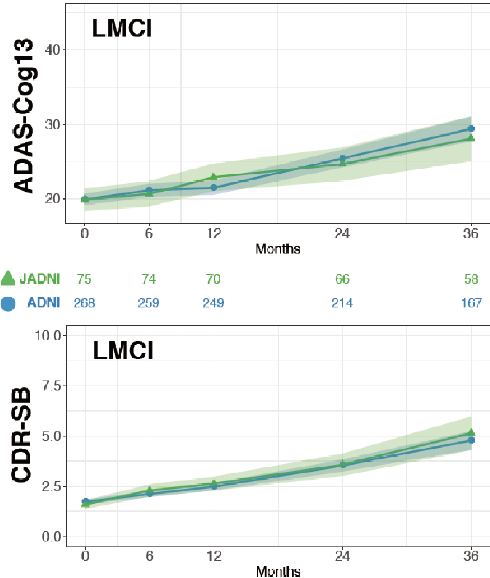
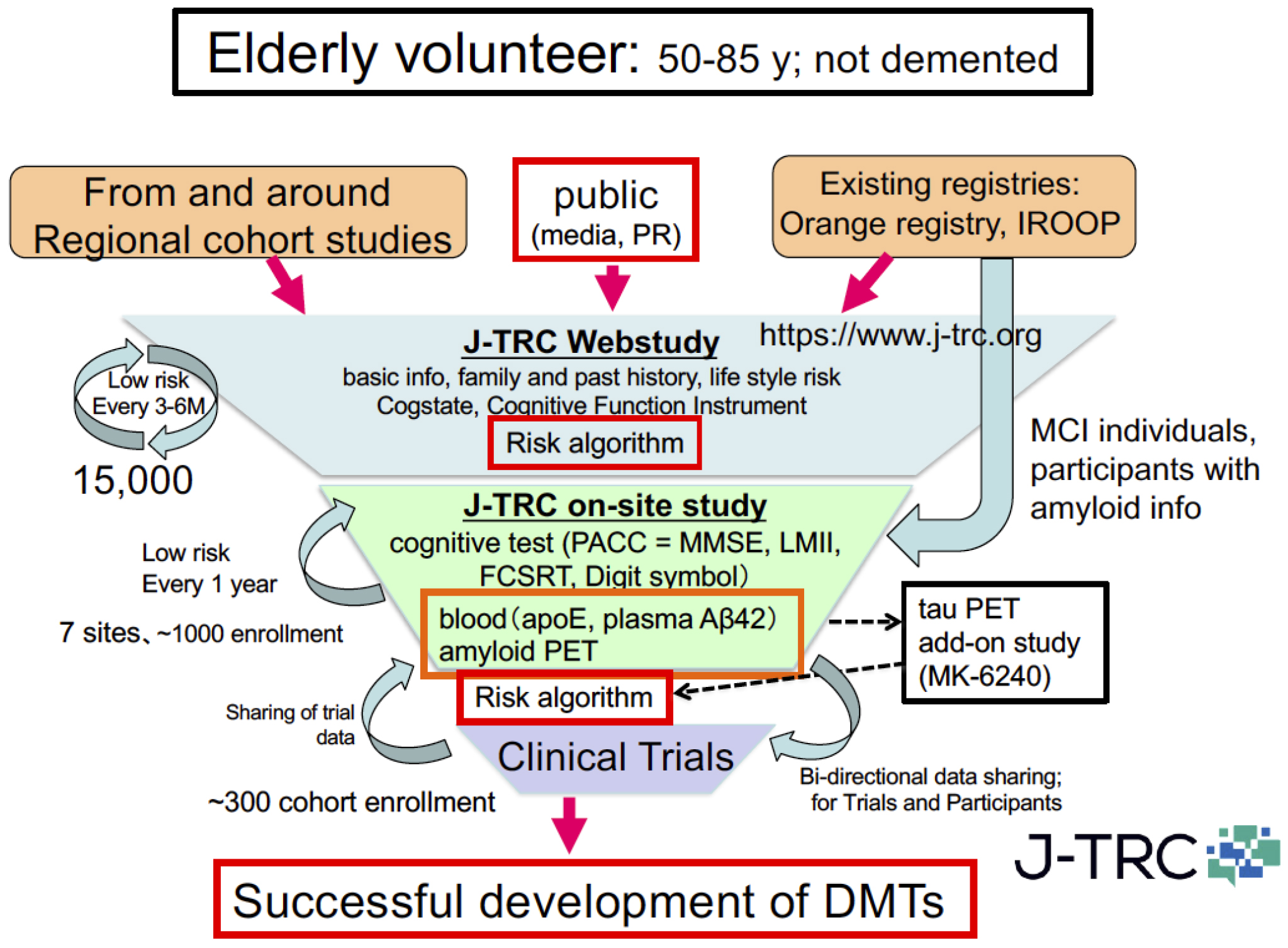
Figure 5. Overview of the Japanese trial-ready cohort (J-TRC) for preclinical and prodromal AD.

From: Molecular Pathogenesis and Disease-modifying Therapies of Alzheimer’s Disease and Related Disorders
PAGE TOP