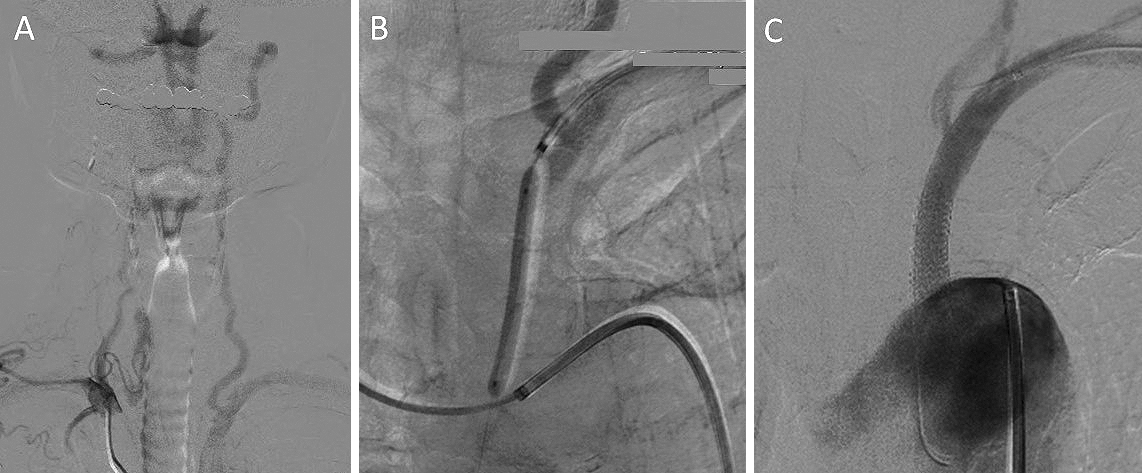
Figure 2. Angiography showed an occlusion at the origin of the left subclavian artery (SA), reversed flow in the left vertebral artery (VA), and identified subclavian steal syndrome (A). Percutaneous transluminal angioplasty with stenting for the proximal left SA occlusion was performed (B), which revealed normal flow in the left VA (C).
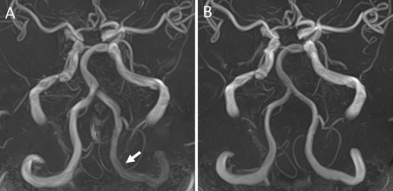
From: Brain Magnetic Resonance Angiography of Subclavian Steal Syndrome