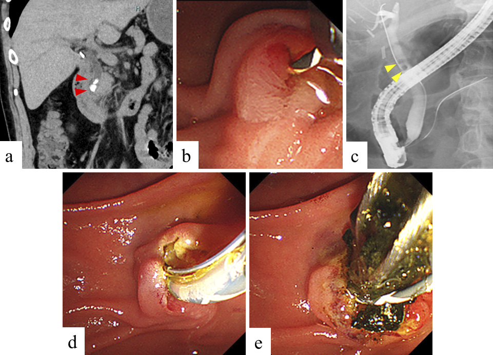
Figure 1. Preprocedural images and images during ERCP in group A.
a. Computed tomography image. A common bile duct stone with a diameter of 5 mm was observed in distal bile duct. (red arrow).
b. Guidewire was inserted into the common bile duct after selective biliary cannulation.
c. Cholangiogram. A common bile duct stone with a diameter of 5 mm was observed in distal bile duct. (yellow arrow).
d. Endoscopic sphincterotomy was performed to extract the stone.
e, f. A common bile duct stone was completely extracted using a basket catheter.
g. No residual stones were observed via cholangiogram using a balloon catheter.
From: Post-endoscopic Retrograde Cholangiopancreatography Pancreatitis after Conservative Treatment for Symptomatic Bile Duct Stones
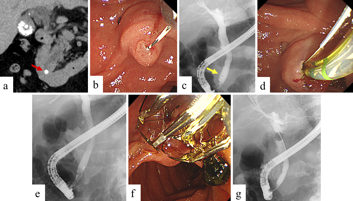
Figure 2. Preprocedural images and images during ERCP in group B.
a. Computed tomography image. Two common bile duct stones with a diameter of 6 mm and 4 mm were observed in distal bile duct. (red arrowheads).
b. Guidewire was inserted into the common bile duct after selective biliary cannulation.
c. Cholangiogram. Two common bile duct stones with a diameter of 6 mm and 4 mm were observed in upper bile duct. (yellow arrowheads).
d. Endoscopic sphincterotomy was performed to extract the stones.
e. Common bile duct stones were completely extracted using a basket catheter.
From: Post-endoscopic Retrograde Cholangiopancreatography Pancreatitis after Conservative Treatment for Symptomatic Bile Duct Stones