Figure 1. Preoperative plain spinal radiographs demonstrated L1 compression fracture (a). T2-weighted imaging on magnetic resonance imaging revealed a recent L1 vertebral compression fracture without spinal canal compromise (b). Plain spinal radiographs after balloon kyphoplasty reveal effective filling of cystic fracture cavity (c).

From: Surgical Treatment of Vertebral Compression Fracture in a Patient with Idiopathic Normal Pressure Hydrocephalus

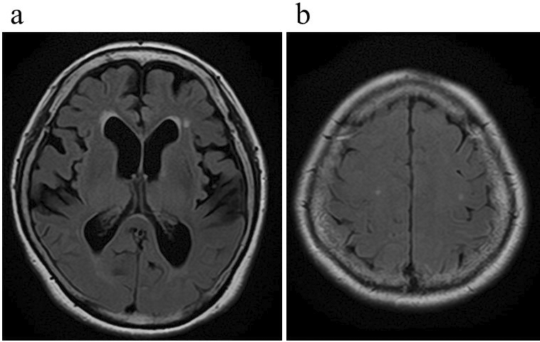
Figure 2. Magnetic resonance imaging showed disproportionately enlarged subarachnoid space hydrocephalus, such as ventriculomegaly with an Evans’ index of 0.35 (a), narrowing of the cerebrospinal fluid spaces near the vertex, and widening of the Sylvian fissure (b).

From: Surgical Treatment of Vertebral Compression Fracture in a Patient with Idiopathic Normal Pressure Hydrocephalus

Figure 3. Postoperative three-dimensional computed tomography showed the programmable valve and catheter’s correct placement.

From: Surgical Treatment of Vertebral Compression Fracture in a Patient with Idiopathic Normal Pressure Hydrocephalus

PAGE TOP