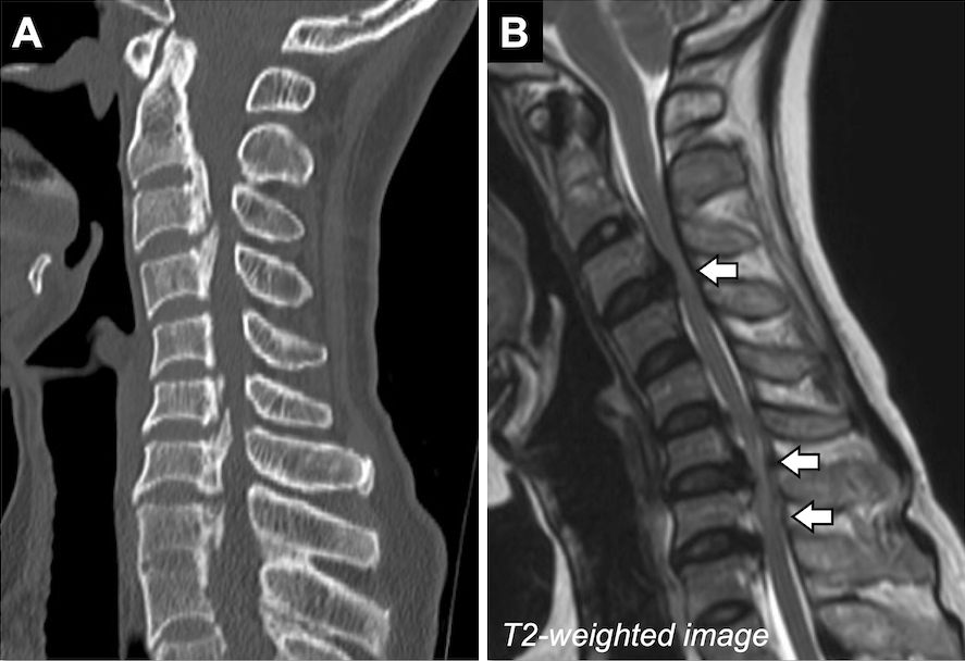
Figure 1. A) Spine computed tomography showed ossification of the posterior longitudinal ligament from C2 to T1. B) Magnetic resonance imaging of the cervical spine revealed intramedullary high signal intensity on T2-weighted images at the C3/4 and C6-T1 levels (arrows).