Figure 1. Dynamic imaging of urine flow at the bladder neck by the air bubble (A, B, C, D, E).
(A) The closed bladder neck before urination; (B) air bubble rotated from 4 o’clock position (C) to 6 o’clock (D) and 11 o’clock position and (E) sucked into the internal urethra until it disappeared.
From: In Vivo Vortex Imaging of Bladder
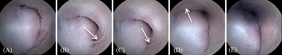
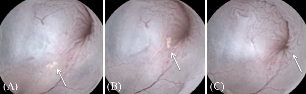
Figure 2. Dynamic imaging of urine flow at the bladder neck by the epithelial cluster (A, B, C).
(A) Appearance of the epithelial cluster (B, C) and its gradual disappearance during voiding.
From: In Vivo Vortex Imaging of Bladder