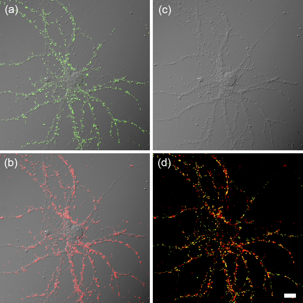
Figure 1. Immunocytochemical labeling of pre- and postsynaptic molecules in cultured hippocampal neurons illustrates the distribution of numerous synaptic connections formed onto branching dendrites. (a) Distribution of the postsynaptic protein, Homer1 (green) overlaid with a differential interference contrast (DIC) image. (b) Distribution of the presynaptic protein, vesicular glutamate transporter 1 (VGLUT1; red) overlaid with a DIC image. (c) DIC image of the same neuron exhibiting the pattern of branching dendrites. (d) Overlaid image of Homer1 (green) and VGLUT1 (red). Bar, 10 μm.
From: Development and Application of Technology for Neural Circuit Visualization - Secondary Publication