Corresponding author: Yoshito Nishimura, me421060@s.okayama-u.ac.jp
DOI: 10.31662/jmaj.2018-0031
Received: September 6, 2018
Accepted: September 26, 2018
Advance Publication: November 12, 2018
Published: March 4, 2019
Cite this article as:
Otsuka Y, Harada K, Nishimura Y, Otsuka F. Ludwig’s Angina and Mandibular Osteomyelitis after Dental Extraction. JMA J. 2019;2(1):87-88.
Key words: Ludwig’s angina, Osteomyelitis, Dental infection, Imaging
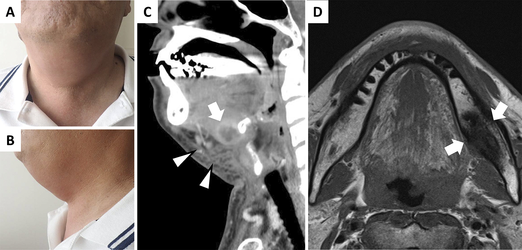
A 63-year-old Japanese man was admitted to our hospital owing to dysphagia, hoarseness, and jaw swelling (Figure 1A, B). He had undergone left lower wisdom-tooth extraction 3-weeks prior to admission. Contrast-enhanced computed tomography (CT) demonstrated submandibular cellulitis and abscess formation (Figure 1C). Magnetic resonance imaging (MRI) showed a low signal intensity lesion in the left mandible on the T1-weighted image (Figure 1D). These findings were consistent with Ludwig’s angina complicated with abscess and submandibular osteomyelitis. Blood cultures obtained on the day of admission showed negative results. Intravenous ampicillin/sulbactam was administered, and the symptoms improved. Ludwig’s angina, a severe cellulitis in submandibular space, is most commonly caused by a dental infection, especially in the second or third molar (1). Submandibular osteomyelitis is a severe complication after dental therapy; however, its diagnosis is often neglected because of its indeterminate symptoms (2). Furthermore, owing to the low sensitivity of CT in the diagnosis of osteomyelitis, MRI is essential when osteomyelitis is suspected.

None
YO wrote the manuscript. KH was an inpatient attending physician and edited the manuscript. YN edited the manuscript. FO supervised all the procedures.
Informed consent was obtained from the patient.
Boscolo-Rizzo P, Da Mosto MC. Submandibular space infection: a potentially lethal infection. Int J Infect Dis. 2009;13(3):327-33.
Pineda C, Espinosa R, Pena A. Radiographic imaging in osteomyelitis: the role of plain radiography, computed tomography, ultrasonography, magnetic resonance imaging, and scintigraphy. Semin Plast Surg. 2009;23(2):80-9.