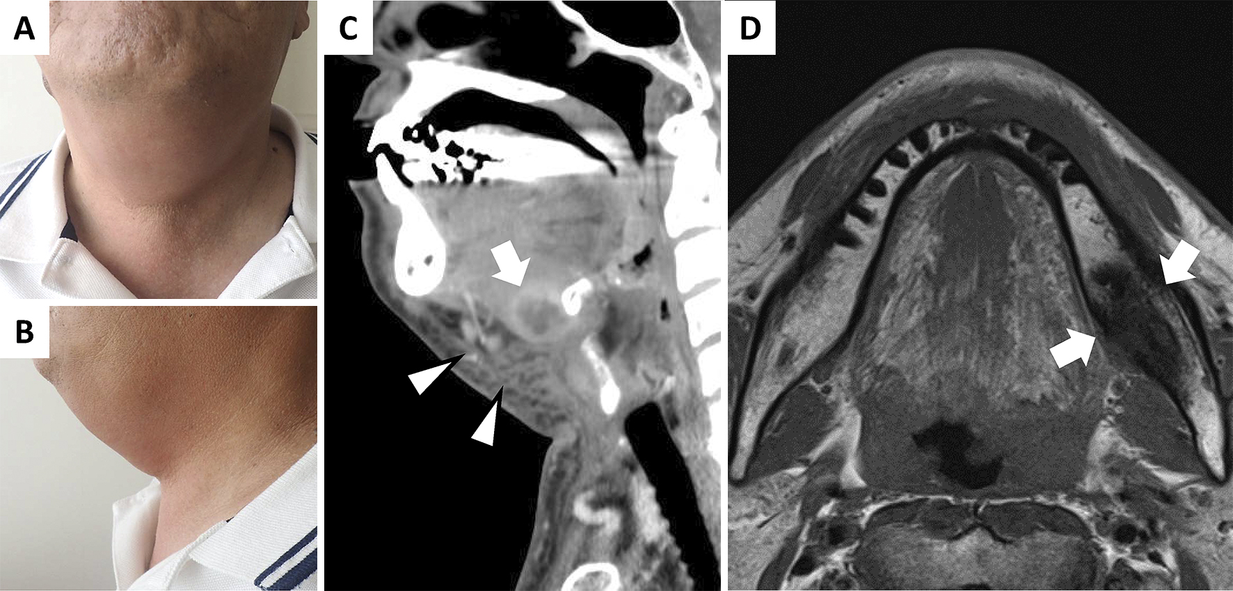
Figure 1. (A), (B) Physical examination revealed swelling and redness in the jaw. (C) Contrast-enhanced computed tomography (CT) demonstrated cellulitis in the patient’s jaw (arrowheads) and formation of an abscess (20-mm diameter) in front of the hyoid bone (arrow). (D) Magnetic resonance imaging (MRI) showed a low signal intensity lesion in the body of the left mandible on the T1-weighted image (arrows).
From: Ludwig’s Angina and Mandibular Osteomyelitis after Dental Extraction

