Corresponding author: Akira Kuriyama, ak13568@kchnet.or.jp
DOI: 10.31662/jmaj.2021-0031
Received: March 16, 2021
Accepted: May 6, 2021
Advance Publication: July 9, 2021
Published: July 15, 2021
Cite this article as:
Kuriyama A. Melanosis Coli. JMA J. 2021;4(3):291-292.
Key words: melanosis, constipation, laxatives, anthraquinone
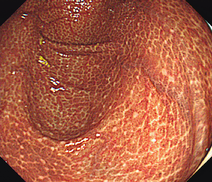
A 75-year-old man with a 23-year history of poorly controlled type 2 diabetes mellitus presented with chronic constipation. Colonoscopy showed heavily pigmented mucosa, resembling leopard skin, from the cecum through the ascending colon (Figure 1). The patient had been using senna glycoside for the last 7 years, which supported a diagnosis of melanosis coli.
A multicenter observational study with patients who had undergone colonoscopy suggested that the prevalence of melanosis coli is approximately 1.8% (1). Melanosis coli is associated with the chronic use of laxatives, particularly those containing anthraquinones, such as senna, rhubarb, and cascara. It can develop within a few months of using anthraquinone-containing laxatives. Anthraquinones cause direct injury to and apoptosis of the colonic epithelial cells, resulting in lipofuscin deposition in the macrophages of the lamina propria (2), which is visible as a dark pigment. Melanosis coli disappears with the discontinuation of the drug.
Although there is no known association between melanosis coli and colorectal cancer (3), (4), melanosis coli may be associated with a higher incidence and number of colonic non-adenoma polyps and low-grade adenomas (1), (5). Thus, follow-up colonoscopy should be considered in patients with melanosis coli.
None
AK took care of the patient, wrote the manuscript, and submitted the current article.
Written informed consent was obtained from the patient to publish this case report including the accompanying images.
Wang S, Wang Z, Peng L, et al. Gender, age, and concomitant diseases of melanosis coli in China: a multicenter study of 6,090 cases. PeerJ. 2018;6:e4483.
Byers RJ, Marsh P, Parkinson D, et al. Melanosis coli is associated with an increase in colonic epithelial apoptosis and not with laxative use. Histopathology. 1997;30(2):160-4.
Biernacka-Wawrzonek D, Stepka M, Tomaszewska A, et al. Melanosis coli in patients with colon cancer. Przeglad gastroenterologiczny. 2017;12(1):22-7.
Kassim SA, Abbas M, Tang W, et al. Retrospective study on melanosis coli as risk factor of colorectal neoplasm: a 3-year colonoscopic finding in Zhuhai Hospital, China. Int J Colorectal Dis. 2020;35(2):213-22.
Liu ZH, Foo DCC, Law WL, et al. Melanosis coli: Harmless pigmentation? A case-control retrospective study of 657 cases. PloS one. 2017;12(10):e0186668.