Corresponding author: Masato Okada, bestlupusdoctorever@gmail.com
DOI: 10.31662/jmaj.2024-0196
Received: August 6, 2024
Accepted: January 31, 2025
Advance Publication: April 4, 2025
Published: April 28, 2025
Cite this article as:
Shiba H, Ozawa H, Okada M. Polymyalgia Rheumatica Presenting as Fever of Unknown Origin. JMA J. 2025;8(2):622-623.
Key words: polymyalgia rheumatica, fever of unknown origin, PET/CT
A 76-year-old woman presented with persistent low-grade fever. She was initially suspected of blood culture-negative infective endocarditis. She underwent an aortic valve replacement and antibiotic treatment. The resected mass was, however, non-infective Lambl’s excrescences. 18-fluorine-fluorodeoxyglucose positron emission tomography/computed tomography (18F-FDG-PET/CT) was performed to detect foci of infection. Instead, it revealed FDG uptake, including around the shoulders and hips, in the interspinous regions and adjacent to the ischial tuberosities (Figure 1). She was referred to the rheumatology department. Focused history-taking revealed bilateral thigh pain with morning stiffness. Physical examination showed edematous extremities and limited range of motion in the wrists and ankles. Ultrasonography identified bilateral biceps tenosynovitis and distal synovitis. Rheumatoid factor and anti-cyclic citrullinated peptide antibodies were negative. She was diagnosed with polymyalgia rheumatica (PMR). Lambl’s excrescences were considered a coincidental finding unrelated to PMR.
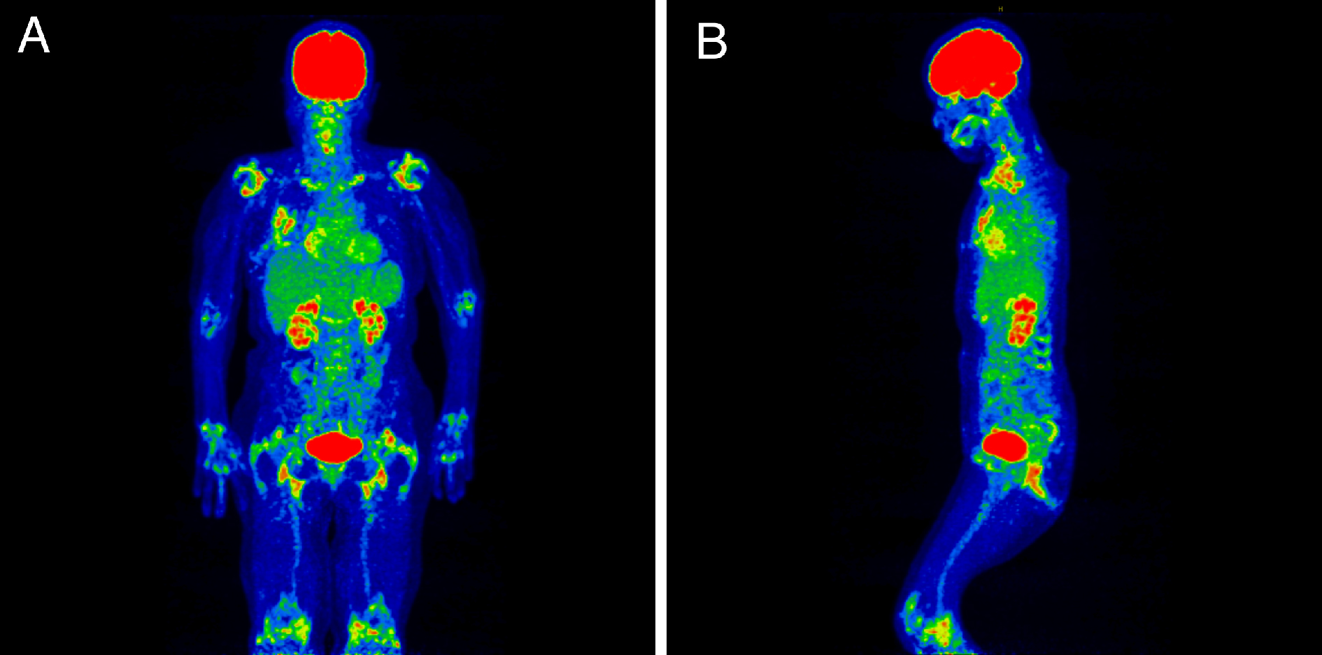
The use of 18F-FDG-PET/CT is currently supported in the diagnostic process of fever of unknown origin (1), (2). Furthermore, this modality has emerged as a gold standard investigation for PMR (3). Its combined pathognomonic PET/CT findings include FDG uptake in the periarticular regions of the shoulders and hips, in the interspinous regions, and adjacent to the ischial tuberosities (3). PET/CT also helps to identify coexistent vasculitis and exclude relevant differential diagnoses including clinically silent extra septic foci (3), (4).
None
HS acquired data and drafted the manuscript. HO and MO reviewed and supervised the manuscript.
We have obtained informed consent for this manuscript.
Wright WF, Kandiah S, Brady R, et al. Nuclear medicine imaging tools in fever of unknown origin: time for a revisit and appropriate use criteria. Clin Infect Dis. 2024;78(5):1148-53.
Hess S, Noriega-Álvarez E, Leccisotti L, et al. EANM consensus document on the use of [18F]FDG PET/CT in fever and inflammation of unknown origin. Eur J Nucl Med Mol Imaging. 2024;51(9):2597-613.
Owen CE, Poon AMT, Liu B, et al. Characterising polymyalgia rheumatica on whole-body 18F-FDG PET/CT: an atlas. Rheumatol Adv Pract. 2024;8(1):rkae003.
Ghanem-Zoubi N. FDG PET/CT in cardiac infection: does it matter? A narrative review. Infect Dis Ther. 2022;11(5):1769-77.
