Corresponding author: Tomoyuki Fujioka, fjokmrad@tmd.ac.jp
DOI: 10.31662/jmaj.2024-0303
Received: October 8, 2024
Accepted: January 14, 2025
Advance Publication: March 14, 2025
Published: April 28, 2025
Cite this article as:
Yagi R, Yamagiwa K, Fujioka T, Tsuchiya J, Asamori T, Tsutsumi T, Tateishi U. A Case of the Monocle Sign in Facial Nerve Palsy Caused by External Auditory Canal Cancer on 18F-fluorodeoxyglucose-positron Emission Tomography/Computed Tomography. JMA J. 2025;8(2):615-616.
Key words: external auditory canal cancer, facial nerve paralysis, PET/CT
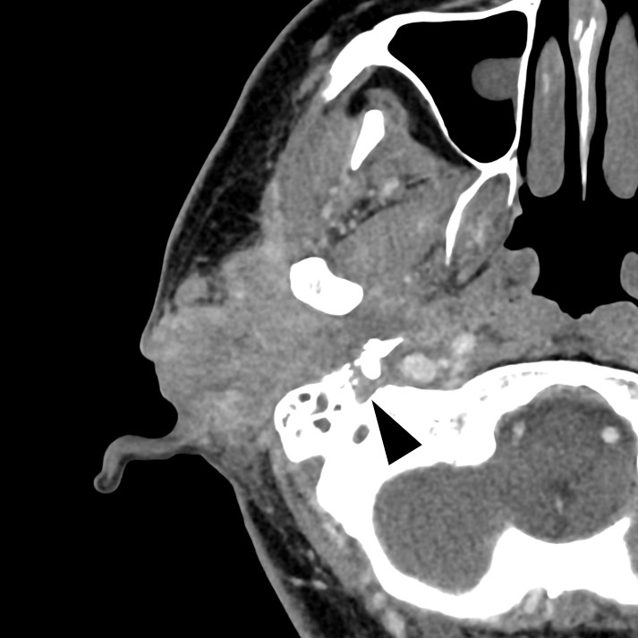
A 47-year-old woman with right external auditory canal cancer presented with an inability to close her left eyelid. Contrast-enhanced computed tomography (CT) identified a tumor extending from the right external auditory canal to the petrous bone, infiltrating the right facial nerve (Figure 1). 18F-fluorodeoxyglucose (18F-FDG)-positron emission tomography (PET)/CT revealed intense 18F-FDG uptake at the site. Notably, the left orbicularis oculi muscle (OOM) showed 18F-FDG uptake (maximum standardized uptake value: 3.08), whereas the right OOM showed no abnormal uptake (Figure 2).
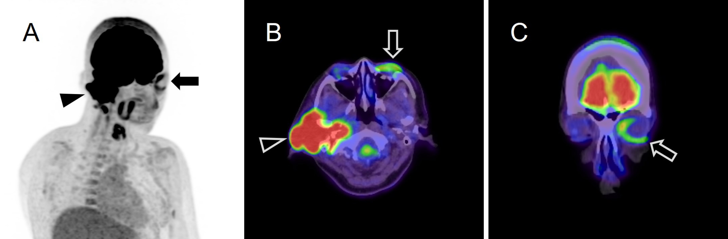
Asymmetrical uptake by the OOM, referred to as the “monocle sign,” indicates contralateral peripheral facial nerve palsy (FNP) (1), which is believed to result from the overactivity of the nonparetic OOM. To the best of our knowledge, this is the first reported case of the monocle sign in a patient with external auditory canal cancer and FNP (1), (2).
Radiologists should recognize the monocle sign to prevent misdiagnosing unilateral ocular uptake under other conditions.
None
We used GPT-4o (https://chatgpt.com/) for Japanese-English translation and English proofreading. The authors read, revised, and proofread the generated text.
Reiko Yagi wrote the first draft of the manuscript, and Tomoyuki Fujioka, Ken Yamagiwa, Junichi Tsuchiya, Tomoaki Asamori, and Takeshi Tsutsumi revised the manuscript. Tomoaki Asamori and Takeshi Tsutsumi contributed to patient care. Ukihide Tateishi supervised all the procedures.
We have obtained informed consent for this case report.
Not applicable.
Orita E, Meerwein CM, Pizzuto DA, et al. The monocle sign in FDG-PET: a sign of contralateral facial nerve palsy. Clin Nucl Med. 2020;45(2):e94-5.
Dana F, Maurer A, Muehlematter UJ, et al. The monocle sign on 18F-FDG PET indicates contralateral peripheral facial nerve palsy. Clin Nucl Med. 2024;49(8):709-14.
